

Ijraset Journal For Research in Applied Science and Engineering Technology
Brain Tumor Detection using Deep Learning
Authors: Adish Shivdikar, Mihir Shirke, Ishwar Vodnala, Prof. Jaychand Upadhaya
DOI Link: https://doi.org/10.22214/ijraset.2022.40710
Certificate: View Certificate
Abstract
Tumors are now the second major cause of cancer. A huge percentage of patients are in danger as more than just a consequence of cancer. The medical field needs fast, automated, efficient and reliable technique to detect tumor like brain tumor. Detection plays very important role in treatment. If proper detection of tumor is possible then doctors keep a patient out of danger. Various image processing techniques are used in this application. Doctors are able to provide excellent treatment and save a huge number of tumour patients by using this application. A tumour is nothing more than an uncontrolled growth of cells. Brain tumour cells expand to the point where they consume all of the nutrition intended for healthy cells and tissues, resulting in brain failure. Currently, doctors manually locate the position and area of a brain tumour by looking at the patient\'s MR images of the brain. This leads to inaccuracy in tumour detection and is extremely time intensive. A tumour is an uncontrollably growing clump of tissue. We can utilise CNN (Convolution Neural Network), also known as NN (Neural Network), and VGG 16 Deep Learning architectures (visual geometry group). To diagnose a brain tumour, transfer learning is used. The model\'s performance predicts whether or not a tumour is present in an image. If a tumour is present, the answer is yes; otherwise, the answer is no.
Introduction
I. INTRODUCTION
A. Brain Tumor Detection System
The brain is the most crucial and vital organ in the human body. A brain tumour is one of the most common causes of brain dysfunction. A tumour is nothing more than an uncontrolled growth of cells. Brain tumour cells expand to the point where they consume all of the nutrition intended for healthy cells and tissues, resulting in brain failure. Currently, clinicians manually locate the position and area of a brain tumour by looking at the patient's MR images of the brain. This leads to inaccuracy in tumour detection and is extremely time intensive. Brain cancer is a deadly disease that claims the lives of many people. A technique for detecting and classifying brain tumours is available, allowing it to be diagnosed at an early stage. Cancer classification is one of the most difficult aspects of clinical diagnosis. This research is about a system that employs computer-based techniques to locate tumour blocks and classify the type of tumour in MRI scans of various patients using the Convolution Neural Network Algorithm. For the detection of brain tumours in MRI scans of cancer patients, several image processing techniques such as image segmentation, image enhancement, and feature extraction are applied. Image Pre-Processing, Image Segmentation, Feature Extraction, and Classification are the four processes involved in detecting a brain tumour utilising image processing technique. The performance of detecting and categorising brain tumours in MRI images is improved using image processing and neural network approaches.
The brain is the most important component of the human neurological system. It is found in the human head and is hidden by the skull. The human brain's job is to coordinate all of the body's functions. It is a sort of organ that enables a person to tolerate and survive a wide range of environmental conditions. The human brain allows people to act and communicate their thoughts and feelings. We will outline the structure of the brain in this section to help you comprehend the fundamentals.
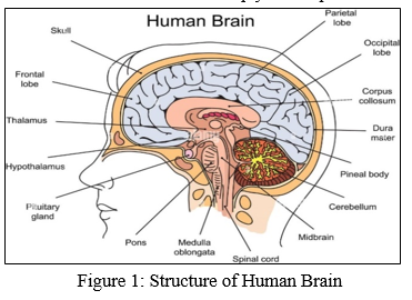
Primary brain tumours (benign tumours) and secondary brain tumours are the two basic forms of brain cancers (malignant tumour). Gliomas are a form of benign tumour in which one type of cell grows slowly in the brain. It comes from astrocytes, which are non-neuronal brain cells. Primary tumours are less aggressive than secondary tumours, but they put a lot of strain on the brain, which causes it to cease working correctly. Secondary tumours are more aggressive and spread into other tissues more quickly. A secondary brain tumour arises from somewhere else in the body. These tumours have a metastatic cancer cell in the body that has spread to other parts of the body, such as the brain and lungs. Secondary brain tumours are extremely dangerous. Lung cancer, kidney cancer, bladder cancer, and other cancers are the most common causes of secondary brain tumours.
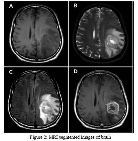
In 1969, Raymond v. Damadian created the first magnetic image. The first MRI image of the human body was created in 1977, and it was the most flawless technique at the time. We can see the many types of tissues in the human body thanks to MRI since it allows us to see the intricacies of the internal structure of the brain. When compared to other medical imaging modalities such as X-ray and computer tomography, MRI images are of higher quality. [8]. MRI is a useful tool for determining the location of a brain tumour in the human body. T1 weighted, T2 weighted, and FLAIR (fluid attenuated inversion recovery) weighted MRI images for mapping tumour caused change are displayed in the picture.
B. Application
The applications' primary goal is to detect tumours. The major goal of this application's creation is to deliver effective treatment as quickly as possible and to protect human lives that are in danger. This application is beneficial to both doctors and patients. Manual identification is slower, but it is more precise and efficient for the user. This application was created to address those issues. It's an easy-to-use programme.
C. Objective
To supply good software to doctors in order for them to diagnose tumours and their causes. To save the time of the patient and to provide a suitable remedy at an early stage. This would make it easier to obtain prompt consultation.
The fundamental goal of brain tumour detection is to be able to not only identify but also classify different types of tumours. It can be useful in situations when we need to know whether a tumour is positive or negative; it can detect tumours from images and return a positive or negative answer. This research is about a system that employs computer-based techniques to locate tumour blocks and classify the type of tumour in MRI scans of various patients using the Convolution Neural Network Algorithm.
II. LITERATURE SURVEY
Paper-1: Brain Tumor Detection and Classification Using Convolutional Neural Network and Deep Neural Network
Publication Year: 2020
Author(s): Chirodip Lodh Choudhury, Chandrakanta Mahanty, Raghvendra Kumar, Brojo Kishore Mishra
Summary: In this research paper, the authors have proposed a new system based on CNN, which discriminates between the Brain MRI images to mark them as tumorous or not. The model achieved an accuracy of 96.08%, with an f-score of 97.3. The model is having CNN with 3 layers and requires very few steps of pre-processing to produce the results in 35 epochs. The purpose of the research is to highlight the importance of diagnostic machine learning applications and predictive treatment.
Paper-2: Brain Tumor Detection Based on Multimodal Information Fusion and Convolutional Neural Network
Publication Year: 2019
Author(s): Ming Li, Lishan Kuang, Shuhua Xu, Zhanguo Sha
Summary: In this paper, a method with three-dimensional MRI brain tumor detection combining multimodal information fusion and CNN is proposed. Firstly, it is better to use the improved multimodal 3D-CNNs to obtain the three-dimensional features of brain tumor lesions under different modalities. The experimental results show that the three evaluation indexes of dice, SN and SE are optimized respectively, and the two-dimensional brain tumor detection network and the original single-mode brain tumor detection method are compared.
Paper-3: Brain Tumor Detection Using Convolutional Neural Network
Publication Year: 2019
Author(s): Tonmoy Hossain, Fairuz Shadmani Shishir, Mohsena Ashraf, MD Abdullah Al Nasim &, Faisal Muhammad Shah
Summary: The paper focuses on detecting brain tumors using machine learning. The authors of this paper had compared the SVM Classification Technique and CNN Classification Technique. So, as per the paper first model is segmented by Fuzzy C Means Algorithm (FCM) and then classified by a traditional machine learning algorithm. The second model focused on deep learning for tumor detection. FCM gives better results for noisy clustered data set. SVM Classification Technique gives 92.42% of accuracy. And 5-layer CNN Classification Technique gives 97.87% of accuracy.
Paper-4: Classification of Brain Tumors from MRI Images Using a Convolutional Neural Network
Publication Year: 2020
Author(s): Milica M. Badža and Marko C. Barjaktarovi´c
Summary: The paper discusses the detection of brain tumors for three types- meningioma, glioma, pituitary tumor. So, they had taken images from three different planes- sagittal plane, axial plane, coronal plane. Then, with all the images Data Augmentation process is performed. Then it goes through a 5-layer CNN classification technique. Further, the output came from the CNN process is been trained by use of Confusion Matrices Algorithm. So,the best result for 10-fold cross-validation was achieved for the record-wise method and, for the augmented dataset, and the accuracy was 96.56%.
Paper-5: Deep Learning Based Image Classification and Abnormalities Analysis of MRI Brain Image.
Publication Year: 2019
Author(s): P.Muthu Krishnammal ,S. Selvakumar Raja
Summary: In this paper, a convolutional neural network (CNN) is designed for classifying the tumor. For extracting quantitative information from an image discrete wavelet transform was used for extracting wavelet coefficients and gray-level co-occurrence matrix (GCLM) for statistical feature extraction. Uses a K -means segmentation algorithm for localizing and segmentation of tumor part once the classification of tumor image was done This study used a dataset of 100 images for training the model and 40 epochs.
Paper-6: Brain Tumor Detection Using Deep Neural Network and Machine Learning Algorithm
Publication Year: 2019
Author(s): Masoumeh Siar, Mohammad Teshnehlab
Summary: In this research paper, a new system which is a combination of clustering algorithm for feature extraction and CNN was proposed. For evaluation purpose, different classifiers such as Softmax Fully Connected layer classifier, RBF classifier and the DT classifier in the CNN architecture was used. This paper uses a dataset that was collected from the imaging center.
III. PROPOSED SYSTEM
The suggested system is composed mostly of five parts. Dataset gathering, pre-processing, and analysis, Split the data, train a Deep Neural Network for epochs, then classify it using a CNN model. We can take many MRI pictures and use one as the input image in a dataset. The image is pre-processed to encode the label and resize it. We divided the data into 80 percent Training Data and 20 percent Testing Data while splitting the data. Then, for each epoch, build a CNN model and train a deep neural network. The image was then classified as yes or no, with yes indicating a positive tumour and no indicating a negative tumour.
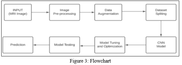
- Dataset collection: Ahmed Hamada published the dataset, which was previously used by numerous researchers in their research papers, on the Kaggle website. There are 3116 MRI pictures in total in this dataset.
- Preprocessing: A preprocessing step is done on the MRI images before feeding them to the proposed structure. In the picture preprocessing section, the keras library's ImageDataGenerator method is used to generate batches of tensor image data with real-time data augmentation so that our model receives a variety of images. The preprocessed input argument of the ImageDataGenerator method of the keras library is also set to an appropriate MRI picture in the format required by the model. This ImageDataGenerator method of the keras library does all essential preprocessing tasks such as cropping, rotation, determining brightness range, flipping the picture, and obtaining the 4 rectangle boundary coordinates of the cropped image.
- Data Modelling: After standardising our complete dataset, we divided it into two sets: a training set and a test set. Data is used for training in 80% of cases and testing in 20% of cases. The photos are then divided into test and training sets using Image Data Generator.
- Methodology: When developing deep learning models, transfer learning is a knowledge-sharing strategy that decreases the bulk of the training data, the time, and the computational expenses. Transfer learning allows a pre-trained model's learning to be transferred to a new model. Tumor classification, software defect prediction, activity recognition, and sentiment classification are just a few of the fields where transfer learning has been employed.
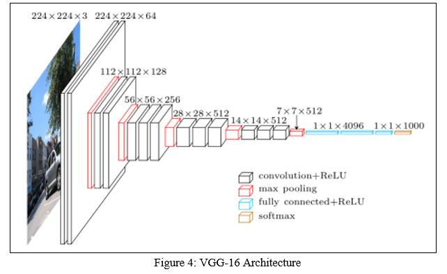
The suggested Deep CNN model was compared to the popular transfer learning approach VGG16 in this study. A convolutional neural network is VGG16. The input to the first convolution layer is a 224 by 224 RGB image with a fixed size. The image is processed through a series of convolutional layers, each having a receptive field of 33 (the smallest size that can capture the concepts of left/right, up/down, and centre). It also uses 11 convolution filters in the configurations, which can be viewed as a linear change of the input channels. The convolution stride is set to 1 pixel, as is the convolution spatial padding.
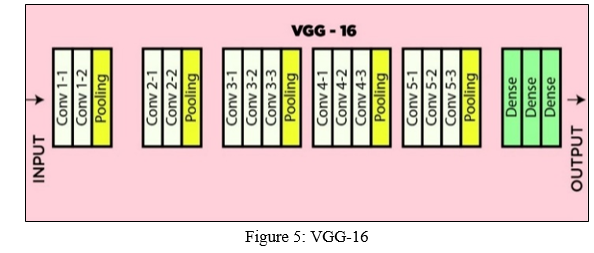
Spatial pooling is performed by five max-pooling layers that are applied after several convolution layers (not all the conv. layers are followed by max-pooling). Maxpooling is done with stride 2 over a 22 pixel window. Three Fully-Connected (FC) layers follow a stack of convolutional layers with varying depths in different designs, the first two of which have 4096 channels each, the third of which conducts 1000-way ILSVRC classification and has 1000 channels, one for each class. The soft-max layer is the final layer. Every network has the same fully linked layer configuration. The rectification (ReLU) nonlinearity is present in all hidden layers. It should also be highlighted that, with the exception of one, none of the networks incorporate Local Response Normalization (LRN), which does not improve performance on the ILSVRC dataset but increases memory consumption and computation time.
Spatial separable and depthwise separable convolutions are the two basic types of separable convolutions. Depthwise Convolution is a type of convolution in which each information channel has just one convolution channel. The channel is almost as deep as the information in typical 2D convolution conducted over several information channels, allowing us to freely combine channels to construct any component in the yield. Depthwise convolutions, on the other hand, keep each channel independent. Multiple filters in a depthwise separable CNN map the input image for distinct patterns. These filters look for patterns one by one and pass them on to the next layer in the architecture. When compared to traditional CNN, it has the advantage of higher accuracy and the ability to recognise several patterns at the same time.
As shown in Figs., the greatest accuracy was between 82 and 92 percent, and the minimum loss was between 0.0017 and 0.5175 with 30 epochs.
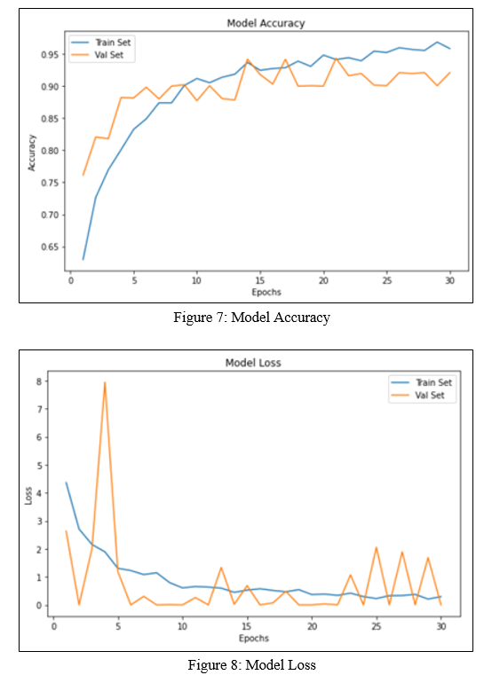
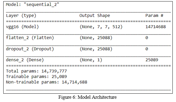
The best performance was attained with the VGG-16, which had a 92 percent accuracy rating. Other algorithms were also reasonably accurate in predicting brain tumours.
Conclusion
In recent years, magnetic resonance imaging (MRI) has proven to be an excellent technique for clinical research, with applications such as brain tumour detection. When applied to these MRI pictures, deep learning algorithms aid in the detection of the tumour. On the MRI image dataset, we applied two techniques: CNN and the VGG-16 model. In comparison to CNN, experimental results demonstrate that VGG-16 provides superior accuracy. The accuracy of the test set utilising VGG-16 was found to be 92 percent. In the healthcare field, the system will be quite beneficial. The proposed approach can determine whether or not a tumour exists. However, in the future, the system could be improved to recognise a specific type of tumour and provide therapy accordingly.
References
[1] Rupal R. Agravat, Mehul S. Raval, “ Prediction of Overall Survival of Brain Tumor Patients “, 2019 IEEE Region 10 Conference (TENCON 2019) 978-1-7281-1895-6. [2] Tonmoy Hossain, Fairuz Shadmani Shishir , Mohsena Ashraf, “Brain Tumor Detection Using Convolutional Neural Network”, 1st International Conference on Advances in Science, Engineering and Robotics Technology 2019 (ICASERT 2019) 978-1-7281-3445-1. [3] P. Mohamed Shakeel, Tarek E. El. Tobely, Haytham Al-feel, Gunasekaran Manogaran, and S. Baskar “ Neural Network Based Brain Tumor Detection Using Wireless Infrared Imaging Sensor”, 2019 IEEE. Translations and content mining are permitted for academic research, ISSN: 2169-3536. [4] Subhashis Banerjee, Francesco Masulli, Sushmita Mitra, Brain Tumor Detection and Classification from Multi-Channel Magnetic resonance imagings using Deep Learning and Transfer Learning”(2017). [5] Sneha Khare, Neelesh Gupta, Vibhanshu Srivastava, “Optimization Technique, Curve Fitting and Machine Learning used to Detect Brain Tumor in Magnetic resonance imaging”, 2014 IEEE International Conference on Computer Communication and Systems(ICCCS 114), Feb 20-21, 2014 Chennai, INDIA. [6] G.Hemanth, M.Janardhan, L.Sujihelen, “Design And Implementing Brain Tumor Detection Using Machine Learning Approach” Proceedings of the Third International”,Proceedings of the Third International Conference on Trends in Electronics and Informatics (ICOEI 2019),ISBN: 978-1-5386-9439-8. [7] Sanjay Kumar, Dr. Ashish Negi, Dr. J.N Singh, Mr. Himanshu Verma, “A Deep Learning for Brain Tumor Magnetic resonance imaging Images Semantic Segmentation Using FCN “, 2018 4th International Conference on Computing Communication and Automation (ICCCA), ISBN: 978-1- 5386-6947-1. [8] Astina Minz, Prof. Chandrakant Mahobiya,” Magnetic resonance imaging Image classification using adaboost for brain tumor type”, 2017 IEEE 7th International Advance Computing Conference (IACC), ISBN: 978-1-5090-1560-3. [9] Komal Sharma, Akwinder Kaur,Shruti Gujral Assistant Professor M.E - CSECU, Gharuan Mohali, Punjab, INDIA , ”Brain Tumor Detection based on Machine Learning Algorithms”, International Journal of Computer Applications (0975 – 8887) Volume 103 – No.1, October 2014 . [10] Fatemeh Derikvand,Hassan Khotanlou,”Brain Tumor Segmentation in Magnetic resonance imaging Images Using a Hybrid Deep Network Based on Patch and Pixel”, 2020 International Conference on Machine Vision and Image Processing (MVIP), ISBN: 978-1-7281-6832-6.
Copyright
Copyright © 2022 Adish Shivdikar, Mihir Shirke, Ishwar Vodnala, Prof. Jaychand Upadhaya. This is an open access article distributed under the Creative Commons Attribution License, which permits unrestricted use, distribution, and reproduction in any medium, provided the original work is properly cited.

Download Paper
Paper Id : IJRASET40710
Publish Date : 2022-03-09
ISSN : 2321-9653
Publisher Name : IJRASET
DOI Link : Click Here
 Submit Paper Online