

Ijraset Journal For Research in Applied Science and Engineering Technology
Survey on Brain Tumor Detection Using Image Processing and Machine Learning Algorithms
Authors: Prof. Akshada Dighe, Vaibhav Pol, Vaishnavi Kulkarni, Harshali Agarwal, Shreeya Sapate, Akshada Shingote
DOI Link: https://doi.org/10.22214/ijraset.2022.41379
Certificate: View Certificate
Abstract
Known to be inherently serious and deadly, a brain tumor is often a life-threatening condition because of its location in the small intracranial cavity (tissue inside the skull). Most research shows that more people in developed countries suffer from brain tumor than in developing countries. In comparison to other methods, this method accurately determines the stage and size of the tumor and identifies the stage of the tumor by analysing the region of the tumor. For this work, k-means and fuzzy c-means are used as algorithms to segment the brain tumor.
Introduction
I. INTRODUCTION
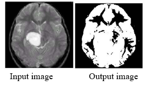
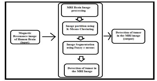
In general, CT scan or MRI that is directed into the intracranial cavity produces a complete image of the brain. After studying several statistical analyses of the people who are affected by brain tumor, some general Risk factors and Symptoms have been identified. The development of technology in science is a day and night process that tries to develop new technologies for treatment. This image is inspected by a physician to detect and diagnose a brain tumor. In the present study, segmentation of brain tumor was performed using the k-means algorithm and fuzzy c-means algorithm to identify tumor stages and sizes. This method accurately determines the stage and size of the tumor. Using this method, tumor tissue can be segmented with a high degree of accuracy and reproducibility comparable to manual segmentation. In addition, the time required to analyze tumor tissue is reduced, and a stage of tumor can be determined from the given area.
Here we have developed an easy to use, cost efficient, fast way of identifying tumor stages using Java.
II. BRAIN MRI IMAGES AS RAW DATA
A tumor is an uncontrolled growth of tissue in any part of the body. It can be primary or secondary. A primary tumor is one that develops at it’s place of origin. A secondary tumor develops from a part of a primary tumor that has spread to a different locations. It normally causes strokes when brain tumor affect CSF (Cerebral Spinal Fluid). This indicates that stroke treatments should be given rather than tumor treatments. Therefore, these treatments require early detection of tumor.
With magnetic resonance image (MRI), one can able to produce detailed images of the inside of the body using strong fields and radio waves.
Most parts of the body can be examined with an MRI scan, including:
- Brain and Spinal Cord.
- Breasts.
- Bones and Joints.
- Internal Organs (liver, womb, etc.)
- Heart and Blood Vessels.
III. ALGORITHMS AND TECHNIQUES
Machine Learning algorithms are usually used in these image processing works as they are clustering algorithms. With the exception of these algorithms, in the previous papers grayscale imaging was done, along with the thresholding method.
A. Segmentation Algorithms
In segmentation algorithms, an image is split into regions or sets of pixels. The purpose of segmentation is to understand better what the image represents. The sets of pixels may represent objects in the image that are of interest to a specific application. Fuzzy C Means is the segmentation algorithms going to be used here. An image data clustering method, fuzzy c-means (FCM), organizes a dataset into N clusters, in which every data point belongs to every cluster to a higher or lower degree.
B. Clustering Algorithms
The clustering algorithm is unsupervised, which means the input is not a labelled one and the problem solving is based on the experience the algorithm gains when solving similar problems as a training schedule K-Means is the clustering algorithm going to used here. As part of the K-means clustering algorithm, centroid points are computed and repeated until the optimal centroid is found. The number of clusters is presumed to be known. This is what flat clustering algorithm is called.
C. Gray Scale Imaging
Grayscale images consist of only one color, meaning they have no color information. Each pixel within a grayscale image represents one of the different gray levels. A standard grayscale image contains 8 bits/pixel data, resulting in 256 gray levels. Digital images are grayscale when the value of each pixel is solely based on the intensity of the light. Such images typically display only the darkest black to the whitest white. In other words, the image contains only black, white, and gray colour.
D. Thresholding Process
With thresholding, we convert a color or grayscale image into a binary image, i.e., one that is simply black and white. Thresholding helps us analyze images better because it not only changes the pixels of an image, but also makes it easier to analyze. This paper concentrates on different clustering-based thresholding algorithms categorized in six groups on the basis of the information they manipulate.
IV. EXISTING SYSTEMS
A typical existing method for detecting malignant tumor relies on thresholding and region growing. In the thresholding method, the spatial characteristics were ignored. Spatial characteristics are essential for malignant tumor detection.
When thresholding-based segmentation is applied, images are considered to have only black or white values. Bit map images contain gray scale values ranging from 0-255. Because of this, sometimes tumor cells are ignored; Additionally, it might not provide an acceptable result for all images because of the intensity inhomogeneity problem.
V. PROCESSING
As needed for the next level, the pre-processing step converts the image. It performs filtering of noise and other artifacts in the image and also sharpens the edges in the image. RGB to gray conversion and reshaping are also taken place here.
The median filter in this paper is used for noise reduction. There is very little possibility of noise arriving in modern magnetic resonance imaging (MRI) scans, but it may occur due to the thermal effect. The aim of this paper is to detect and segment the tumor cells.
Conclusion
In this paper, we have designed an application which uses Magnetic Resonance Image of an human brain to detect the tumor in it. Not necessary that all the images should contain tumor in it. This proposed system processes the image which undergoes through the various clustering and segmentation techniques which helps to detect the exact location of tumor. And by using arithmetic algorithms can detect the size of tumor and is mentioned in the report. After the successful detection, the stage of tumor is to be predicted as if at high risk, low risk or moderate risk. And at the end a report will be generated by the application side. That is all we have concluded from it.
References
[1] S.Mary Praveena ,Dr.I1aVennila , June 2010, \"Optimization Fusion Approach for [mage Segmentation Using K-Means Algorithm,\" International Journal of Computer Applications (0975 - 8887) Volume 2 - NO.7. [2] Manisha Bhagwatl, R.K.Krishna& V.E.Pise July-December 2010, \"Image Segmentation by Improved Watershed Transformation in Programming Environment MATLAB,\" International Journal of Computer Science & Communication Vol. I, No. 2, pp. 17/-/74. [3] K.S. Ravichandran and 2B. Ananthi(2009), \"Color Skin Segmentation Using K-Means Cluster,\" International Journal of Computational and Applied Mathematics ISSN 1819-4966 Volume 4 Number 2,pp. 153 -157. [4] T. Kanungo, D. M. Mount, N. Netanyahu, C. Piatko, R. Silverman, & A. Y.Wu (2002) , \"An efficient k-means clustering algorithm: Analysis and implementation\", Proc. JEEE Con! Computer Vision and Pattern Recognition, pp.881-892.
Copyright
Copyright © 2022 Prof. Akshada Dighe, Vaibhav Pol, Vaishnavi Kulkarni, Harshali Agarwal, Shreeya Sapate, Akshada Shingote. This is an open access article distributed under the Creative Commons Attribution License, which permits unrestricted use, distribution, and reproduction in any medium, provided the original work is properly cited.

Download Paper
Paper Id : IJRASET41379
Publish Date : 2022-04-11
ISSN : 2321-9653
Publisher Name : IJRASET
DOI Link : Click Here
 Submit Paper Online