

Ijraset Journal For Research in Applied Science and Engineering Technology
COVID-19 Detection using Chest X-Ray
Authors: Nirmiti Sali, Nimisha Khadilkar, Srushti Biwalkar, Amisha Swamy, Nusrat Ansari
DOI Link: https://doi.org/10.22214/ijraset.2022.41814
Certificate: View Certificate
Abstract
Over the past few months, the exponential increase in COVID-19 cases has been overwhelming for many healthcare systems across the world. With 114 million cases globally as of 28th February 2021, with India itself having 11.1 million cases, it has challenged us with the testing, quarantine, and safety measures. Having limited testing kits, not all patients that have symptoms of respiratory illness can be tested using conventional techniques (RT-PCR). In this project, we propose the use of chest X-Ray to prioritize the selection of patients for further RT-PCR testing. It would also help in identifying patients with a high likelihood of COVID with a false negative RT-PCR who would wish to repeat testing. Further, we propose the utilization of recent AI techniques to detect the COVID-19 patients automatically using X-Ray images, particularly in settings where radiologists aren\'t available, and help make the proposed testing technology scalable.
Introduction
I. INTRODUCTION
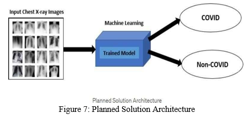
The sudden spike in the number of patients with COVID-19, a new respiratory virus, has put an unprecedented load on healthcare systems across the world. In many countries, healthcare systems have already been overwhelmed. As there are limited kits available, for diagnosis, along with limited hospital beds for admission of such patients, and limited personal protective equipment (PPE) for healthcare personnel, it is thus very important to differentiate which patients with severe acute respiratory illness (SARI) could have COVID-19 infection to efficiently utilize the limited resources. In this work, we propose the use of chest X-Ray to detect COVID-19 infection in patients exhibiting symptoms of SARI. Our tool can classify a given X-Ray as one among the three classes: normal, pneumonia, and COVID pneumonia. The main contribution of this work is in proposing a unique deep neural network-based model for highly accurate detection of COVID-19 infection from the chest XRay images of the patients. Therefore this automated tool can serve as a guide for those at the forefront of this analysis. As we are seeing now too, (February 2021), despite many vaccines in use and development, our country is facing a second wave of the virus and the virus is spreading again. To combat this, our system will provide faster results and help in detection and prevention of further spread faster.
We have also considered symptoms to know whether covid is symptomatic or not.For this we have used Decision tree Algorithm.
II. LITERATURE REVIEW
A. Respiratory illness detection in Chest X-Rays
Various deep learning-based approaches square measure developed to identify completely different diseases like respiratory illness [7, 8, 11, 13]. The model is trained to classify X-Ray pictures into fourteen completely different sickness classes, as well as respiratory illness. Seeing the similarity of the input samples, we tend to found this to be the nearest pre-trained backbone to develop a model for characteristic COVID-19 respiratory illness.
B. COVID-19 detection in Chest X-Rays
Their square measure solely restricted such ASCII text file applications obtainable to be used [1,5,10] that use chest X-Ray pictures. COVID-Net [10] has AN ASCII text file and actively maintained tool which can determine COVID-19 moreover as different respiratory illness whereas showing respectable sensitivity for COVID-19 detection.
[14]have given a fast, absolutely parameterizable GPU implementation of Convolutional Neural Network variants. This paper explains the basics of a system of logic and its use for image classification. It uses Matlab’s system of logic tool cabinet at intervals the definition of a system of logic illation rules. These rules square measure tested and verified through the simulation of classification procedure indiscriminately sample areas. [15]
III. IMPLEMENTATION DETAILS
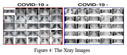
We have used the COVID chest x-ray dataset for COVID-19 frontal-view chest X-Ray images and chest x-ray pneumonia dataset for frontal-view chest X-Ray images with bacterial/viral pneumonia as well as of normal lungs. We will use the pre-trained CNN model, thus implicitly using robust features obtained after training on the Chest X-ray dataset. The two types of images in the obtained dataset are normal and pneumonia. The images in the Normal category belong to the Non-Covid case. But not all the images in Pneumonia come under the Covid category; only images in which the chest is affected due to the SARS-CoV-2 virus are referred to as COVID cases. This is because COVID-19 pneumonia is a subset of pneumonia diseases. The dimensions of all images are not fixed and had to be resized to 100 x100 pixels for training and evaluation. The details about the downloaded dataset are shown below.
|
Sr no. |
Set |
Normal |
Pneumonia |
Covid-19 +ve |
Total |
|
1. |
Training |
1266 |
3418 |
460 |
5144 |
|
2. |
Testing |
317 |
855 |
116 |
1288 |
Figure 1: Composition of Kaggle Dataset
As the dataset was obtained in a raw form wherein the images are classified in two different folders i.e. training and testing, the major challenge we faced was to prepare a single dataset that contains the images of all the three categories (Normal, Pneumonia, and Covid +ve) and the corresponding category of every image. This was achieved by combining the images present in three different folders into one folder and adequately renaming the images by the category they belong in so that images can be differentiated from each other.
A. Pre-Processing
The images are resized or scaled right down to 100x100 size and converted to grey scale. After resizing, on flattening all the images’ arrays, the entire no. of features is 145200 which might cross the available RAM size while training the model on this dataset leading to a session crash. to beat this, we applied a feature reduction technique called Histogram of Oriented Gradients (HOG) which reduced the feature size of images to a good extent i.e., 24336 features. So, in this way, we obtained the reduced feature size of every image within the dataset.
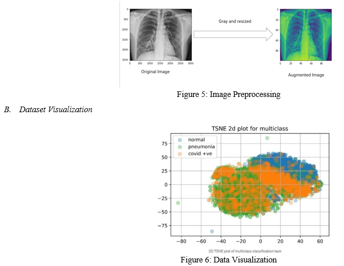
After plotting both the classification tasks using the 2D -distributed stochastic neighbor embedding (TSNE), we conclude that the knowledge isn't fully linearly distributed, there is a big amount of nonlinearity associated with the dataset.
For the multiclass classification problem, the quantity of samples belonging to COVID +ve is identical to the binary classification task. The other two classes are. NORMAL and PNEUMONIA, each containing 1266 and 3418 samples respectively.
Binary Classification task is performed for Detecting covid with help
of symptoms. Decision Tree is a Supervised learning technique that can be used for both classification and Regression problems, but mostly it is preferred for solving Classification problems. It is a tree-structured classifier, where internal nodes represent the features of a dataset, branches represent the decision rules and each leaf node represents the outcome.
C. Methodology
- Libraries to be Used: Matplotlib,Numpy,os,cv2,keras:Sequential M Conv2D,Activation,Maxpooling2D,etc.
- Data Augmentation: Size: Data is resized to 100x100.More the image size,more is the accuracy as the number of pixels increase.
a. Color: Data is converted from one colour space to another,for eg. gray. As raw data does not have uniformity,we use resizing and recolouring.
b. Intensity: Mostly intensity of pixels of gray scale images have values from 0 to 255.So, we convert pixel intensity values between 0 to 1 by dividing it by 255.
Data Augmentation helps to bring Uniformity in all images thus improving the accuracy.
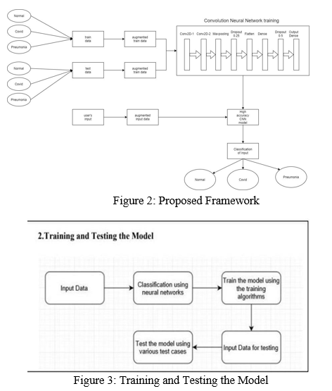
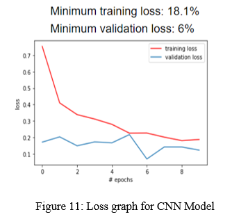
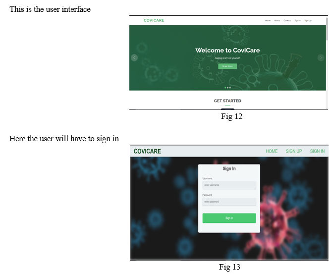
3. Create CNN Model: We need to provide augmented data to CNN model.Deep CNN Model is created using keras Library Dense, Dropout, Flatten, Conv2D,Maxpooling2D -these layers are used. These layers will fetch parameters. Activation ReLU (Rectified Linear Unit) speeds up training and is faster to compute as it brings non linearity. Maxpooling reduces dimensions and computation. Accuracy is calculated by training model with the help of train data.
4. Input From User: The input images also undergo augmentation and then they are passed through CNN mode
5. Classification of Input: Input Data is processed in the model and the class to which it belongs i.e. Covid,Normal or Pneumonia is given as output.
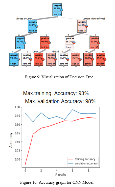
For detection of COVID from symptoms, the parameters like cough, fever, age,etc. are considered and classified using Decision Tree Algorithm. Decision tree helps to predict class of given symptoms i.e Covid Positive or Negative
This algorithm compares the values of root attribute with the record (real dataset) attribute and, based on the comparison, follows the branch and jumps to the next node until it reaches the leaf node of the tree.
IV. EXPERIMENTAL RESULTS
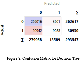
A. Binary Classification Task (Covid +ve vs Covid -ve)
The symptoms gives the result as covid +ve or -ve using Decision Tree algorithm. The confusion matrix for the same is shown here:
V. APPLICATION
Looking at the current situation, the most viable and efficient solution is that this could be used to ramp up the testing with no delays and treatment could be started with immediate effect.
A. Comparison with Other Tests
Antigen test - Not accurate, nowhere accepted to get hospitalized, requires raw material and not accessible everywhere
RT PCR – Accepted (At Least 2days result delay) , but you also need to attach HRCT scan to get admitted which is expensive
Our project – Accurate enough plus no further test needed, can be used to Understand seriousness with no time delay, Cost efficient. No raw material required and almost available everywhere.
This is the user interface
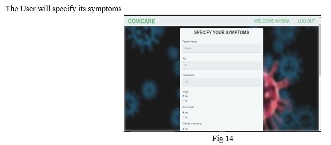
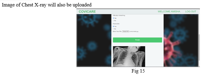
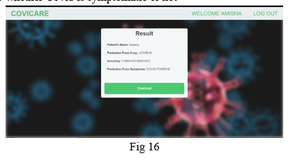
On clicking the Predict button the result will appear on the screen. The final result will be given by Image of Chest X-ray. The result given by symptoms is only to know whether Covid is symptomatic or not
VI. FUTURE SCOPE
To create a csv file from recorded data for convenience in using data.
To use Location to keep a record of spread of Covid in a particular region.
This will help the doctors to study the relation between symptoms and Chest X ray.
VII. ACKNOWLEDGEMENT
We wish to express a true sense of gratitude towards our Mentor Mrs. Nusrat Ansari for giving us this opportunity to work on this project, using our skills and knowledge. We would like to extend our gratitude to all team members for their time-to-time support and hard work because of which we can build a well-written paper. With all respect and gratitude, we appreciate our success to the writers of reference papers that are referred to by us in completion of this paperwork activity which will be useful in presenting our survey paper.
Conclusion
As we know the detection of Covid-19 from chest X-ray images is very important for both doctors and patients to decrease the diagnostic time, reduce financial costs and be able to save more lives. For recognizing images for the tasks thought, artificial intelligence and deep learning are capable. As we have seen, several experiments were performed for the high-accuracy detection of COVID-19 in chest X-ray images. When the number of images in the database and the detection time of COVID-19 (average testing time = 0.03s/image) are considered, it can be suggested that the considered architectures reduce the computational cost with high performance. We got the results that showed the convolutional neural network with minimized convolutional and fully connected layers is capable of detecting COVID-19 images within the two-class, COVID19/Normal and COVID-19/Pneumonia classifications with a mean accuracy of 85%. Our project is a humble venture to satisfy the needs of common citizens for efficient detection of COVID-19. In this study, we used CNN model for detection of Covid From Chest X ray and Decision for detection from Symptoms. This project is aimed at helping doctors in making decisions in their clinical practice due to its high performance and effectiveness, this also gives an insight to how CNN was used to automatically detect the COVID-19.
References
[1] Bennett, J.M.: Smart ct scan based covid19 virus detector. https://github.com/JordanMicahBennett/SMART-CT-SCAN_BASED-COVID19_VIRUS_DETECTOR [2] Cohen, J.P., Morrison, P., Dao, L.: Covid-19 image data collection. arXiv2003.11597 (2020), https://github.com/ieee8023/covid-chestxray-dataset [3] Huang, G., Liu, Z., van der Maaten, L., Weinberger, K.Q.: Densely connected convolutional networks. In: Proceedings of the IEEE Conference on Computer Vision and Pattern Recognition (2017) [4] Mooney, P.: Kaggle chest x-ray images (pneumonia) dataset. https://www.kaggle.com/paultimothymooney/chest- xray-pneumonia (2018) [5] Nisar, Z.: https://github.com/zeeshannisar/covid-19. https://github.com/zeeshannisar/COVID-19 (2020) [6] Petsiuk, V., Das, A., Saenko, K.: Rise: Randomized input sampling for the explanation of black-box models. In: Proceedings of the British Machine Vision Conference (BMVC) (2018) [7] Rajpurkar, P., Irvin, J., Zhu, K., Yang, B., Mehta, H., Duan, T., Ding, D., Bagul, A., Langlotz, C., Shpanskaya, K., et al.: Chexnet: Radiologist-level pneumonia detection on chest x-rays with deep learning. arXiv preprint arXiv:1711.05225 (2017) [8] Ranjan, E., Paul, S., Kapoor, S., Kar, A., Sethuraman, R., Sheet, D.: Jointly learning convolutional representations to compress radiological images and classify thoracic diseases in the compressed domain (12 2018). https://doi.org/10.1145/3293353.3293408 [9] Ruiz, P.: Understanding and visualizing densenets. https://towardsdatascience.com/understanding-and- visualizing-densenets-7f688092391a (2018) [10] Wang, L., Wong, A.: Covid-net: A tailored deep convolutional neural network design for detection of covid- 19 cases from chest radiography images (2020) [11] Wang, X., Peng, Y., Lu, L., Lu, Z., Bagheri, M., Summers, R.M.: Chestx-ray8:Hospital-scale chest x-ray database and benchmarks on weakly-supervised classification and localization of common thorax diseases. In: Proceedings of the IEEE conference on computer vision and pattern recognition. pp. 2097–2106 (2017) [12] Weng, X., Zhuang, N., Tian, J., Liu, Y.: Chexnet for classification and localization of thoracic diseases. https://github.com/arnoweng/CheXNet/ (2017) [13] Yao, L., Poblenz, E., Dagunts, D., Covington, B., Bernard, D., Lyman, K.: Learning to diagnose from scratch by exploiting dependencies among labels. arXiv preprint arXiv:1710.10501 (2017)
Copyright
Copyright © 2022 Nirmiti Sali, Nimisha Khadilkar, Srushti Biwalkar, Amisha Swamy, Nusrat Ansari. This is an open access article distributed under the Creative Commons Attribution License, which permits unrestricted use, distribution, and reproduction in any medium, provided the original work is properly cited.

Download Paper
Paper Id : IJRASET41814
Publish Date : 2022-04-24
ISSN : 2321-9653
Publisher Name : IJRASET
DOI Link : Click Here
 Submit Paper Online