

Ijraset Journal For Research in Applied Science and Engineering Technology
A Comprehensive Review on Detecting Diabetic Eye Diseases Using Deep Learning and Machine Learning Models
Authors: Sudha Abirami R, Suresh Kumar G
DOI Link: https://doi.org/10.22214/ijraset.2023.55596
Certificate: View Certificate
Abstract
Diabetic Ocular Diseases (DOD), or Diabetic Eye Diseases (DED), is a set of eye problems that can affect people with all types of diabetes. Severe diabetes without proper diagnosis and control may lead to vision loss. It might damage the optical nerve, which causes poor/blurry vision or blindness. The problem includes Diabetic Retinopathy, Diabetic Macular Edema (DME), Glaucoma, and Cataracts. Diabetes people may frequently have this ailment, manifesting as fuzzy vision, floaters or streaks resembling cobwebs, retinal edema, impaired color perception, and eventually blindness. The primary issue is that these conditions are irreversible. So, timely diagnosis and treatments are a must. Many technologies, particularly Deep Learning (DL) and Machine Learning (ML), have evolved to predict and detect diseases earlier. This study aims to review some previously developed frameworks proposed by authors. Finding a reliable way to detect diseases early is the central stimulation behind this review. Most earlier works focused on categorizing images according to the severity of conditions. However, this review of the classification of diabetic eye diseases may give a proper path to identify suitable CNN for further work.
Introduction
I. INTRODUCTION
A. Diabetic Ocular Diseases
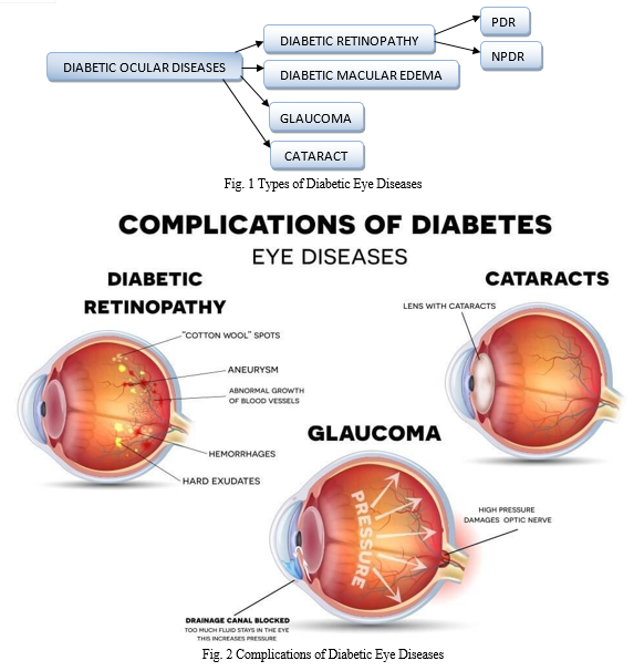
Eye diseases caused by diabetes are due to the high blood sugar level of the patients. Due to its clumsy and asymptotic nature, it is easier to identify if it makes our vision clear. Some kinds of diabetic eye diseases are shown in Fig 1.
- Diabetic Retinopathy: Diabetic retinopathy is a diabetes complication that affects the eyes [as shown in Fig 2]. It affects blood vessels in the retina (the light-sensitive layer of tissue in the back of the eye). At first, diabetic retinopathy might cause no symptoms or only mild vision problems. But it can lead to blindness. The condition can develop in anyone with Type 1 (or) 2 people with diabetes. If the disease worsens, some blood vessels close off, which causes new blood vessels to grow or proliferate on the retina's surface. This stage is called proliferative diabetic retinopathy.
- Diabetic Macular Edema: The part of the retina needed for reading, driving, and seeing faces is called the macula. Diabetes can cause macular edema. Macular edema happens when fluid builds up on the retina and causes swelling and blurry vision. Diabetic macular edema can lead to permanent vision loss.
- Glaucoma: Glaucoma is a group of diseases that cause damage to the optic nerve. This damage leads to irreversible loss of vision. Having diabetes doubles the chance of getting glaucoma.
- Cataract: Excess blood sugar from diabetes can cause cataracts. One may need to undergo cataract surgery to remove lenses that are clouded by the effects of diabetes. Maintaining reasonable control of blood sugar helps prevent permanent clouding of the lens and surgery. [1]
Diabetic Retinopathy has some more types based on severity. They are,
- Non-Proliferative Diabetic Retinopathy- NPDR (Mild, Moderate, and Severe)
- Proliferative Diabetic Retinopathy – PDR
B. Deep Learning
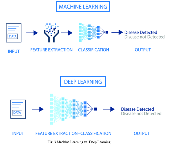
Artificial neural networks, a class of algorithms used in deep learning, are models for the structure and operation of the brain. In other words, it replicates how human brains work. Deep learning algorithms are modeled after how the nervous system is organized, with each neuron communicating with the others.
Deep Learning Algorithms use a neural network to find associations between inputs and outputs. A typical deep learning model has at least three layers and operates in layers. A neural network's information, hidden, and output layers are all made up of “nodes.” Input layers accept data represented numerically (for example, pixel-perfect photographs), output layers produce predictions, and hidden layers handle most of the calculation. Each layer receives the data from the one below and transfers it to the one above.
The feature extraction area is one of the areas where machine learning and deep learning models diverge [refer to Fig 3]. A person performs feature extraction in machine learning, whereas deep learning models come to their conclusions.
C. Machine Learning
Machine learning (ML) is a subset of Artificial Intelligence that uses data as input. Predetermined mathematical functions can yield results with the help of classification or regression techniques. For example, using ML, locating malignant cells in a microscopic image is more straightforward when compared to analyzing it by looking at the pictures. Alzheimer’s disease, Heart Disease, Breast and Lung cancer, and Pneumonia are some diseases that may be identified with ML. The emergence of machine learning (ML) algorithms in disease diagnosis domains illustrates the technology’s utility in medical fields. In addition, suggestions over medicines and diagnosis can also be performed with the help of ML.
Tasks in machine learning are typically categorized into broad groups. These classifications are based on how information is absorbed or how feedback on learning is provided to the system that produced them. The two most popular machine learning techniques are unsupervised learning and supervised learning; both train algorithms using examples of input and output data that humans have labeled. But unsupervised learning gives the algorithm no labeled data to let it discover structure in the input data.
D. Disease Diagnosis
- Diagnosis of Eye Diseases: Diabetic retinopathy can be diagnosed by a comprehensive dilated eye exam. An eye drop has been placed to dilate the pupils, which helps the doctor examine inside our eyes. The bubbles can cause our close vision to blur until they wear off several hours later. During the exam, an eye doctor will look for abnormalities in our eyes’ inside and outside parts. If we have mild or moderate nonproliferative diabetic retinopathy, we might not immediately need treatment. When diabetic retinopathy is mild or moderate, reasonable blood sugar control can usually slow the progression.
- Fluorescein angiography: After our eyes are dilated, a dye is injected into a vein in your arm. The pictures are taken as the dye circulates through our eyes’ blood vessels. The images can pinpoint blood vessels that are closed, broken, or leaking. The healthcare provider will take pictures of the inside of the eye. After the first group of photos is taken, a dye called Fluorescein is injected into a vein. Most often, it is injected inside our elbow. A camera-like device takes pictures as the paint moves through the blood vessels in the back of your eye. A newer method called ultra-wide field Fluorescein Angiography can provide more information about specific diseases than regular angiography [3].
- Optical coherence tomography (OCT): Optical Coherence Tomography is a non-invasive diagnostic technique that renders a vivo cross-sectional view of the retina. OCT utilizes a concept known as interferometry to create a cross-sectional map of the retina that is accurate to within at least 10-15 microns. With this test, pictures provide cross-sectional images of the retina that show the thickness of the retina. This will help determine how much fluid, if any, has leaked into retinal tissue. Later, OCT exams can be used to monitor how the treatment is working.
OCT is useful in diagnosing many retinal conditions, especially when the media is transparent. In general, lesions in the macula are more accessible to the image than in the mid and far periphery. OCT can be beneficial in diagnosing: Macular holes, Macular pucker/epiretinal membrane, Vitreomacular traction, Macular edema, and exudates, Detachments of the neurosensory retina, Detachments of the retinal pigment epithelium (e.g., central serous retinopathy or age-related macular degeneration), Retinoschisis, Pachychoroid, Choroidal tumors. [2]
II. RELATED WORK
Dolly Das et al. [4] reviewed the diagnosis of diabetic retinopathy using machine learning and deep learning. DL models can process smaller datasets using efficient data processing techniques. However, large datasets are generally integrated into deep architectures to improve feature extraction and image classification performance. The author listed some challenges while developing deep learning models. Some of them are feature classification, such as exudates and optic discs, and segmentation of retinal vessels should be appropriately classified. Image preprocessing and data augmentation can be performed to overcome data imbalance through proper sampling. They also stated that, among various methodologies, it was realized that ML techniques are highly scalable concerning high dimensional data and take more time in the analysis and training of models when compared to DL models.
Raffaele Nuzzi et al. [5] stated that A black box suggests a lack of understanding of the algorithm’s decision-making process that gives a specific output. Several techniques are used to bound this phenomenon, such as the “occlusion test, in which a blank area is systemically moved across the entire image and the largest drop in predictive probability represents the specific area of highest importance for the algorithm” or “saliency maps (heat maps) generation techniques such as activation mapping, which, again, highlights areas of importance for classification decisions within an image.”
Srikanta Kumar Padhy et al. [6] carried over a review and found that Machine learning processes mainly include two parts, a training set followed by a validation set. This process occurs by providing a large number of training data, i.e., thousands of retinal images of varying grades of DR to the machine/system as the training set. The authoritative professionals label most of the data as per features in advance. After being exposed to multiple annotated retinal images, the machine learns to grade DR by itself by building a model of complex relationships between input data and generalizing a performance standard. In addition, other data are used to verify the established algorithm, i.e., the validation set. The FDA approval of the Idx-DR device was based on a study on 900?subjects in a primary care setting (10 primary care sites) with automated image analysis. Two 45-degree digital images per eye (one centered on the macula, one centered on the optic nerve) were obtained and analyzed. These images were compared with the stereo, wide-field fundus imaging interpreted by the Wisconsin Fundus Photograph Reading Centre (FPRC). After procuring retinal photos, the artificial intelligence system can make a diagnosis in just 20 seconds.
Shreya Shekar et al. [7] reviewed and analyzed that the collection of retinal datasets is briefly outlined, and then DL methods are discussed. After that, various approaches have been adopted to identify retinal irregularity, including retinal blood vessels, HEMs, Mas, and Exs. Then the performance evaluation metrics were briefly reviewed for automated detection models. The report examined that almost a scholarly job has been done by utilizing CNN models to produce deep multilevel models for detecting DR employing digital retinal photographs. The report examined that almost an academic career has been carried out using CNN models to create deep multilevel models for detecting DR hiring digital retinal photos.
The upcoming chapters are arranged in a manner; in Chapter 3, research works conducted so far on diabetic eye diseases using deep learning and machine learning; in Chapter 4, discussions regarding the results obtained by various researchers; and in Chapter 5, the conclusion.
III. DEEP LEARNING AND MACHINE LEARNING APPROACHES – REVIEW
In this chapter, let us briefly see how deep learning and machine learning approaches influenced the field of Disease Diagnosis shown in Table 1. This table lists all the previously conducted experiments and the results obtained by various authors for identifying and classifying diabetic eye diseases.
Table 1 DL and ML Approaches and their Results
|
AUTHOR |
YEAR |
DATA SET USED |
ML OR DL MODEL |
FEATURES |
RESULTS |
|
[8] |
2022 |
Private Dataset, Messidor-1 and Messidor-2 |
EfficientDet-d1, EfficientDet-d1 End-to-End Deep Fusion model |
Diabetic Macular Edema and Hard Exudates Detection |
This fusion model aimed to detect DME using an Efficient Net-based object classifier and detector as a dual model. Also compared, the results of both double and fusion models with hyperparameter characteristics. |
|
[9] |
2020 |
DR Detection [2015], APTOS 2019 |
DenseNet-169 |
Diabetic Retinopathy severity classes |
This proposed model is trained based on DenseNet-169. This model classifies Diabetic Retinopathy severity cases alone. The results are compared with the regression model and other ML classifiers such as SVM, Decision Tree, and KNN. |
|
[10] |
2021 |
SD-OCT |
Deep Transfer Learning based on Inception v4 |
DR, AMD, DME |
In fine tune ConvNet configuration, the model achieved 96.6% accuracy, whereas, with fixed feature extractor configuration, the model achieved 81%. |
|
[11] |
2020 |
EyePACS (American), Keio (Japanese) |
Support Vector Machine, Deep CNN, and Inception v3 |
DR |
DR Classification model using AI was created based on American fundus images. SVM exhibits superior performance compared with NN architecture. |
|
[12]
|
2021 |
Kaggle, DIARETDB, Messidor |
Faster RCNN+ CNN |
DR lesions, EX and HM lesion ROI |
In the first stage, lesions were detected with regional faster RCNN, and in the second stage, transfer learning and attention mechanism were used for DR classification grading. |
|
[13] |
2021 |
EyePACS, Messidor-1 and Messidor-2 |
Inception ResNet v2 |
DR Grading |
They developed a deep learning agent using Inception ResNetv2 as the base model. Training and testing data sets are different. Additionally, the validation data set was used to evaluate the hyperparameters. |
|
[14] |
2021 |
Private Dataset – Mansoura University, Egypt |
Multifractal+ SVM |
NPDR |
This approach analyzed macular optical coherence tomography angiography (OCTA) images based on Multifractal geometry. SVM with a Multifractal geometry algorithm achieved 98.5% accuracy. |
|
[15] |
2021 |
Kaggle Dataset |
Deep Ensemble Learning and Attention Mechanism |
DR |
Constructed a detection model DR-IIXRN consisting of Inceptionv3, InceptionResnetv2 Xception, ResNeXt101, and NASNetLarge. I also modified the model using transfer learning, fine-tuning, and attention mechanisms to detect DR in an improved manner. The weighted voting algorithm was used to categorize the severity classes. |
|
[16] |
2021 |
EyePACS, DIARETDB0, DIARETDB1, eOphtha, MESSIDOR, DRIVE |
SMOTE, Deep Learning, NASNET-Large |
DR |
This study uses a 2-stage training method to solve the overfitting problem. In the training phase, Learning Module 1 was used to identify the DR and no-DR to build the model. The Learning Module 2 on SMOTE synthetic datasets to identify the mild-NPDR, moderate NPDR, severe NPDR, and proliferative DR classification. |
|
[17] |
2021 |
Private Dataset |
Deep Learning, ResNet |
DR |
The deep DR system consisted of three sub-networks: The image quality assessment sub-network, the lesion-aware subnetwork, and the DR grading sub-network. This system was trained based on ResNet. Retinal images were fed as input to perform the DR classification task. |
|
[18] |
2022 |
Private dataset, Open-source Dataset |
CNN, VGG16 VGG 19 |
DR Severity classes |
This paper presents an automated classification system that analyzes fundus images with varying illumination and fields of view and generates a severity grade for diabetic retinopathy (DR) using machine learning models such as CNN, VGG-16, and VGG-19. This system achieves 80% sensitivity, 82% accuracy, 82% specificity, and 0.904 AUC for classifying images into five categories ranging from 0 to 4, where 0 is no DR, and 4 is proliferative DR. |
|
[19] |
2021 |
APTOS-2019, IDRiD Dataset |
DenseNet-100 |
DR and DME |
DenseNet-100 was used as a feature extraction method on which the one-stage detector, Center Net, is employed to localize and classify the disease lesions. We evaluated our method over challenging datasets, namely, APTOS-2019 and IDRiD, and attained average accuracy of 97.93% and 98.10%, respectively. We also performed cross-dataset validation with benchmark EYEPACS and DIARETDB1 datasets. |
|
[20] |
2022 |
DRISHTI-GS, DRIONS-DB, HRF, PSGIMSR |
ResNet, VGG Net, GoogLeNet |
Glaucoma |
In this proposed work, an ensemble model was designed to detect glaucoma in the early stage. The performance of the proposed method of ensemble architecture is compared with three ConvNet architectures, such as ResNet-50, VGGNet-16, and Google Net, and analyzed with five different data sets. |
|
[21] |
2021 |
Messidor, Messidor-2, DRISHTI-GS, and Retinal Dataset from GitHub |
ResNet50, VGG-16, and Xception |
DR, DME, Glaucoma |
In this research, we are using CNN-based transfer learning to implement the DED retinal fundus image classification. The three pre-trained models, namely; Xception, VGG16, DenseNet21, and the five-layered convolutional model, have been evaluated regarding test data set accuracy. |
|
[22] |
2020 |
APTOS-2019 |
DenseNet121 |
DR |
In this study, we have utilized a pre-trained DenseNet121 network with several modifications and trained on APTOS 2019 dataset. The proposed method outperformed other state-of-the-art networks in early-stage detection. It achieved 96.51% accuracy in severity grading of Diabetic Retinopathy for multi-label classification and performed 94.44% accuracy for the single-class classification method. |
|
[23] |
2021 |
Kaggle |
Deep Learning Based Algorithm |
DR |
CNN Architecture results in 95% using tiny lesions, pre-trained convolutional neural network (CNN) results in 93.37% using some lesions, and LCD Net results 96.5 % to 99.7% accuracy depicts the highest results among all by using optic disk, retinal vasculature, macula, and fovea type features. |
|
[24] |
2020 |
Messidor-2, IDRiD |
EfficientNet-B5 |
DR |
First, the model is trained on a mixture of two Messidor-2 and IDRiD datasets and evaluated on the Messidor dataset. As the pre-processing step, a helpful technique called CLAHE is presented for amplifying the vessels in retinal fundus images. The IDRiD test set includes 516 images that are classified into segregated groups from 0 (No DR) to 4 (Severe DR) |
|
[25] |
2022 |
EyePACS-1, Messidor-2, and DIARETDB0 |
U-Net Model, Transfer Learning based VGGNet |
DR |
In the first stage, we employed two distinct U-Net models for optic disc (OD) and blood vessel (BV) segmentation during the preprocessing. In the second stage, the enhanced retinal images after OD and BV extraction are used as an input of transfer learning-based model VGGNet, which performs DR detection by identifying retinal biomarkers such as Exudates (EX), Microaneurysms (MA), and Hemorrhages (HM). The proposed model achieved state-of-the-art performance with an average accuracy of 96.60%, 93.95%, 92.25% evaluated on EyePACS-1, Messidor-2, and DIARETDB0, respectively. |
|
[26] |
2023 |
BDDR- [IDRiD, EyePACS, APTOS] Dataset |
Blended Deep Learning Model [VGG16/19, ResNet 50/101, InceptionV3, Inception ResNet, Xception, DenseNet, Dark Net & Efficient Net] |
DR |
The authors proposed a blended approach obtained by training several models in this work. We blended the results of ten state-of-the-art deep learning models for DR grading at different resolutions for final scoring. Since we have used 5-fold cross-validation, this gives 50 models. |
|
[27] |
2023 |
APTOS Dataset |
Inception-V3 |
DR |
An automatic DR classification model was developed with two different scenarios: case1, in which the preprocessing step is performed using CLAHE followed by ESRGAN is used, and case2, in which neither step is performed while using augmentation of the images to prevent overfitting in both scenarios. Lastly, images were sent into the Inception-V3 model for the classification step. |
IV. DISCUSSION
According to the review, among the numerous types of diabetic eye problems, most research has been done to identify Diabetic Retinopathy (DR). We can determine the existence of disease based on traits like Optic Disc detection, Exudates detection, Hemorrhages detection, and Micro aneurysms present in blood vessels. The researchers selected various publicly accessible datasets such as EyePACS, Messidor-1 and Messidor-2, APTOS-2019, IDRiD, and private datasets are few of them. Deep Learning and CNN models have often been used techniques for disease classification. Initially, specific image preprocessing techniques are used on the images in the dataset, including pixel transformations, green channel extraction, denoising of the pictures, applying some contrast enhancement, and histogram equalization. Following preprocessing, a training set of data was fed to previously developed suggested models, and the effectiveness of the models was assessed using a test set of data. Support Vector Machine algorithm is also used to predict the disease severity [11], [14]. The authors developed new algorithms from pre-trained models to identify the disease severity classes. In most of the research works it is clearly stated that transfer learning [10], [12], [15], [21], and [25] was applied to improve the performance of the model. And in some cases, DenseNet [9], [19], and [22] played a vital role in classifying the disease cases. At the same time, few research works have been published on ensemble learning techniques [15]. In terms of hyperparameters, most of the proposed works achieved better accuracy, specificity, and sensitivity. Even though the proposed models outperform well in their classification, still Diabetic Retinopathy (DR) only be identified primarily. From all of the previous research works, it is observed that not only the type of Diabetic Retinopathy alone enough but also the detection of all kinds of Diabetic Eye Diseases are essential.
V. ACKNOWLEDGEMENT
We acknowledge Pondicherry University for providing all the resources to write a research paper.
Conclusion
It is observed from the summary table shown in Table 1 and discussions that most of the previous works concentrated on classifying DR only. Even though more powerful commercial AI solutions are available for eye disease screening, not everyone can afford such expensive methods. Therefore, it would be advantageous to detect and categorize all diabetic eye illnesses using a computer-aided disease diagnostic model. Also, opting for deep learning-based models is always better when dealing with a large volume of data. So, an efficient and reliable methodology is required to categorize all Diabetic Eye Diseases (i.e., All-in-One DL Method). Before classification, images may be processed pixel by pixel with augmentation and segmentation techniques. As a result, segmented images simplify the classification task of identifying the severity and kind of diseases. From this classification, accuracy may be improved too. An automated segmentation approach can be proposed to classify medical images to yield better outcomes. This can also be helpful in real-time scenarios in terms of predicting the multi-class classification approach of diabetic eye disease precisely.
References
[1] https://www.niddk.nih.gov/health-information/diabetes/overview/preventing-problems/ [2] https://invisioncare.com/diabetic-eye-disease/ [3] https://medlineplus.gov/ency/article/003846.html [4] Dolly Das, Saroj Kr. Biswas & Sivaji Bandyopadhyay, “A critical review on the diagnosis of diabetic retinopathy using machine learning and deep learning,” Multimedia Tools and Applications Volume 81, pages25613–25655 (2022)”, https://doi.org/10.1007/s11042-022-12642-4. [5] Raffaele Nuzzi, Giacomo Boscia, Paola Marolo and Federico Ricard, “The Impact of Artificial Intelligence and Deep Learning in Eye Diseases: A Review”, Doi: 10.3389/fmed.2021.710329. [6] Srikanta Kumar Padhy, Brijesh Takkar, Rohan Chawla, Atul Kumar, “Artificial intelligence in diabetic retinopathy: A natural step to the future,” doi 10.4103/ijo.ijo _1989_18. [7] Shreya Shekar, Nitin Satpute, and Aditya Guptaa, “Review on diabetic retinopathy with deep learning methods,”https://www.spiedigitallibrary.org/journals /Journal-of-Medical-Imaging on 09 Jan 2022. [8] Ting-Yuan Wang1, Yi-Hao Chen2, Jiann-Torng Chen2, Jung-Tzu Liu1, Po-Yi Wu1†, Sung-Yen Chang1†, Ya-Wen Lee1†, Kuo-Chen Su3 and Ching- Long Chen2 *, “Diabetic Macular Edema Detection Using End-to-End Deep Fusion Model and Anatomical Landmark Visualization on an Edge Computing Device,” https://doi.org/10.3389/fmed.2022.85164. [9] Gazala Mushtaq 1 and Farheen Siddiqui 2, “Detection of diabetic retinopathy using deep learning methodology,” doi:10.1088/1757-899X/1070/1/012049. [10] Fouzia Nawaz1, Muhammad Ramzan1, Khalid Mehmood1, Hikmat Ullah Khan2, Saleem Hayat Khan3,4, and Muhammad Raheel Bhutta5, “Early Detection of Diabetic Retinopathy Using Machine Intelligence through Deep Transfer and Representational Learning,” doi:10.32604/cmc.2020.012887. [11] Yusaku Katada a,b,1, Nobuhiro Ozawa a,b,1, Kanato Masayoshi a, Yoshiko Ofuji b, Kazuo Tsubota b, Toshihide Kurihara, “Automatic screening for diabetic retinopathy in interracial fundus images using artificial intelligence,” https://doi.org/10.1016/j.ibmed.2020.100024. [12] Abdüssamed Erciyas and Necaattin Bar??ç?, “An Effective Method for Detecting and Classifying Diabetic Retinopathy Lesions Based on Deep Learning”, https://doi.org/10.1155/2021/9928899. [13] Gaurav Saxena, Dhirendra Kumar Verma, Amit Paraye, Alpana Rajan, Anil Rawat, “Improved and robust deep learning agent for preliminary detection of diabetic retinopathy using public datasets,” https://doi.org/10.1016/j.ibmed.2020.100022. [14] Mohamed M. Abdelsalam 1, M. A. Zahran, “A Novel Approach of Diabetic Retinopathy Early Detection Based on Multifractal Geometry Analysis for OCTA Macular Images Using Support Vector Machine,” Digital Object Identifier 10.1109/ACCESS.2021.3054743. [15] Zhuang Ai 1†, Xuan Huang2,3†, Yuan Fan1, Jing Feng2, Fanxin Zeng4, and Yaping Lu1, “DR-IIXRN: Detection Algorithm of Diabetic Retinopathy Based on Deep Ensemble Learning and Attention Mechanism,” https://doi.org/10.3389/fninf.2021.778552. [16] Ping Nan Chen1, Chia Chiang Lee2, Chang Min Liang3, Shu I Pao3, Ke Hao Huang3 and Ke Feng Lin2,4, “General deep learning model for detecting diabetic retinopathy,” https://doi.org/10.1186/s12859-021-04005-x. [17] Ling Dai1,2,3,9, Liang Wu 2,9, Huating Li 2,9, Chun Cai 2,9, Qiang Wu4,9, Hongyu Kong 4, Ruhan Liu 1,3, Xiangning Wang4, Xuhong Hou2, Yuexing Liu2, Xiaoxue Long 2, Yang Wen 1,3, Lina Lu5, Yaxin Shen 1,3, Yan Chen4, Dinggang Shen 6,7, Xiaokang Yang8, Haidong Zou 5?, Bin Sheng 1,3? & Weiping Jia, “A deep learning system for detecting diabetic retinopathy across the disease spectrum,” https://doi.org/10.1038/s41467-021-23458-5. [18] Anumol Sajan, Anamika K, Ms. Simy Mary Kurian, “Diabetic Retinopathy Detection using Deep Learning,” International Journal of Engineering Research & Technology (IJERT) ISSN: 2278-0181 Published by, www.ijert.org ICCIDT - 2022 Conference Proceedings. [19] Tahira Nazir 1, Marriam Nawaz 1, Junaid Rashid 2, Rabbia Mahum 1, Momina Masood 1, Awais Mehmood 1, Farooq Ali 1, Jungeun Kim 2, Hyuk-Yoon Kwon 3, and Amir Hussain 4, “Detection of Diabetic Eye Disease from Retinal Images Using a Deep Learning Based CenterNet Model,” https://doi.org/10.3390/s21165283. [20] Shubham Joshi1, B. Partibane,2 Wesam Atef Hatamleh,3 Hussam Tarazi,4 Chandra Shekhar Yadav, five and Daniel Krah, “Glaucoma Detection Using Image Processing and Supervised Learning for Classification,” https://doi.org/10.1155/2022/2988262. [21] Rubina Sarki, Khandakar Ahmed, Hua Wang, Yanchun Zhang, Jiangang Ma, Kate Wang, “Image Preprocessing in Classification and Identification of Diabetic Eye Diseases,” https://doi.org/10.1007/s41019-021-00167-z. [22] Saket S. Chaturvedi, Kajol Gupta, Vaishali Ninawe, Prakash S. Prasad, “Automated Diabetic Retinopathy Grading using Deep Convolutional Neural Network,” https://arxiv.org/ftp/arxiv/papers/2004/2004.06334.pdf. [23] Amita Meshram, Dr. Deepak Dembla, Dr. Reema Ajmera, “Analysis and Design of Deep Learning Algorithms for Retinal Image Classification for Early Detection of Diabetic Retinopathy,” Turkish Journal of Computer and Mathematics Education, Research Article, Vol.12 No.6 (2021), 2633-2641. [24] Pour AM, Seyedarabi H, Jahromi SHA, Javadzadeh A, “Automatic detection and monitoring of diabetic retinopathy using efficient convolutional neural networks and contrast limited adaptive histogram equalization,” IEEE Access 8:136668–13667310.1109/ACCESS.2020.3005044. [25] Anas Bilal, Guangmin Sun, Sarah Mazhar, Azhar Imran & Jahanzaib Latif, “A Transfer Learning and U-Net-based Automatic detection of diabetic retinopathy from fundus images,” https://doi.org/10.1080/21681163.2021.2021111. [26] Fernando C. Monteiro, “Diabetic Retinopathy Grading using Blended Deep Learning,” https://doi.org/10.1016/j.procs.2023.01.389. [27] Ghadah Alwakid, WalaaGouda, and MamoonaHumayun, “Deep Learning-Based Prediction of Diabetic Retinopathy Using CLAHE and ESRGAN for Enhancement”, https://doi.org/10.3390/healthcare11060863.
Copyright
Copyright © 2023 Sudha Abirami R, Suresh Kumar G. This is an open access article distributed under the Creative Commons Attribution License, which permits unrestricted use, distribution, and reproduction in any medium, provided the original work is properly cited.

Download Paper
Paper Id : IJRASET55596
Publish Date : 2023-09-01
ISSN : 2321-9653
Publisher Name : IJRASET
DOI Link : Click Here
 Submit Paper Online