

Ijraset Journal For Research in Applied Science and Engineering Technology
Detection of Leukemia Disease Using Image Processing and Machine Learning
Authors: V. V. Nagamani, Sirigiri Lakshman Sai, Puram Pavan, Chilukuru Venkata Sai
DOI Link: https://doi.org/10.22214/ijraset.2024.60399
Certificate: View Certificate
Abstract
: This paper presents a project that addresses Detection Of Leukemia Disease Using Image Processing and Machine Learning. This research proposes an approach that combines image processing techniques with machine learning algorithms to detect leukemia in microscopic blood smear images at a stage. The proposed method involves enhancing the images through preprocessing identifying the regions of interest through segmentation and extracting features using image processing algorithms. Afterward a machine learning model trained on a dataset of annotated images is employed to classify the samples as either positive or negative, for leukemia. The effectiveness of this approach is evaluated using metrics like accuracy, sensitivity and specificity. Experimental results show promising outcomes in detecting leukemia which could serve as a tool for healthcare professionals, in early diagnosis and treatment planning. By integrating this framework into practice it has the potential to improve efficiency and accuracy in diagnosing leukemia while ultimately leading to patient outcomes and enhanced healthcare management.
Introduction
I. INTRODUCTION
Leukemia is the abnormal proliferation of white blood cells in blood which requires prompt detection and treatment. Traditional methods of diagnosis rely on the manual examination of microscopic blood smears by expert pathologists. This tradition is fraught with challenges such as being highly laborious, time consuming, and prone to subjectivity. In this paper, we propose an integrated methodology, which combines image processing and machine learning for automated leukemia identification. From these sophisticated algorithms, quantitative features are extracted, and machine learning is applied for effective categorization. The proposed framework attempts to improve diagnosis precision and expedite the process.
The contribution of this paper is to illustrate the potential of the integration to redefine leukemia diagnosis. We have analyzed existing literature and our methodology is systematically presented. The primary aim of research is to automate the analysis of blood smear images. We believe this research is of significant interest to clinicians and the general reader. Furthermore, it could become an invaluable tool to automate the diagnosis of leukemia.
II. LITERATURE REVIEW
- Joshi et al. (2013) reported the usage of Otsu’s global thresholding on an enhanced greyscale image. To differentiate blasts in a microscopic blood smear image, they extracted the area, perimeter, and circularity from the equivalent binary image and employed the K-nearest neighbor decision algorithm for classification.
- Moradiamin et al. (2015) converted images from the RGB color space to HSV, which reduced the correlation between the color channels in comparison to RGB and enabled the three H, S, and V channels to be dealt with separately.The dimensionality of the feature set was reduced by principal component analysis some features, which were subsequently applied to the classifier.
- A different approach was introduced by Kazemi et al. (2016) by implementing selective median filtering in combination with conversion to the CIEL∗a∗b model, in which the perceptual difference between colors is proportional to the Cartesian distance. In simple terms, the formula CIEL∗a∗b takes the XYZ tristimulus values and the white reference to produce correlates to the luminence, chroma, and hue elements (Fairchild, 2005).
- Wang et al. (2008) proposed a segmentation algorithm that combined adaptive thresholding with an edge-based technique and seeded watershed to recognize cell nuclei in different cycle phases.
III. PROPOSED SYSTEM
The proposed leukemia detection approach enhances cell identification by combining image processing and machine learning. It outperforms conventional approaches by utilizing strong algorithms that assess cellular features in pictures.
This union enhances diagnostic precision, which is crucial for leukemia detection and treatment planning.The advantages of this proposed system include:
- Increased Accuracy: Through combining advanced image processing and machine learning algorithms, the system identifies subtle cellular features that are indicative of leukemia, leading to improved diagnostic accuracy over current methods.
- Early Detection: The system has the potential to detect leukemia in patients well before it may be diagnosed with current methods, allowing for earlier treatment and improved outcomes
- Objective Analysis: The system provides an objective analysis of cellular features, reducing the reliance upon subjective human interpretation. This consistency is significantly enhancing the reliability of leukemia diagnosis and prognosis.
- Efficiency: Automated analysis of cell images reduces the time taken to reach a diagnosis, allowing healthcare professionals to optimally utilize their time and resources.
- Personalized Treatment Planning: By accurately identifying leukemia cells, the method we depict can assist in the development of leukemia treatment plans by enabling clinicians to develop highly personalized treatment plans for patients based on specific characteristics of the disease.
- Scalable: We believe our system will be scalable because the integration of image processing and machine learning algorithms work well in handling many data, as such we believe our system may be suitable for widespread implementation across healthcare facilities.
- Continuous Learning: The system's machine learning models can continuously learn from new data, enabling the system to improve its performance over time and keep up with the latest advancements in leukemia detection.
IV. METHODOLOGY
The goal is to create an image processing system for blood cell counting, which is essential in leukemia identification. Leukemia is typically accompanied with a drop in white blood cell count, which influences illness incidence. The white blood cell to red blood cell ratio is helpful in prognosis.
The strategy, which emphasizes affordability, helps hematologists forecast results. Previous research developed a low-cost image processing tool for this purpose. The technique involves using this equipment to perform precise cell count analysis in order to detect leukemia. The following steps outline our methodology:
- Data Collection and Preprocessing: Gather a variety of blood cell pictures including both healthy and unhealthy samples. Clean and preprocess these images to clarify the features and reduce noise, making for the best input for analysis.
- Feature Extraction: Extract the relevant features from the preprocessed images. These might include cell size, shape, and color, among others.
- Algorithm Selection: When selecting image processing algorithms to use for cell counting, keep in mind concerns such as efficiency and accuracy.
- System Design: Make a plan for the design of your image processing system, including the algorithms that you'll use to perform the processing and making sure that the system will be able to scale.
- Development: Using the most appropriate programming languages and frameworks for the task, create the system ensuring that the implementation is interoperable with a number of devices.
- Testing and validation: Take different datasets to test the accuracy, reliability and the performance of the system.
- Maintenance and updates: A system has to be maintained for optimal working and to update it time to time for bugs and performance issues and changed requirements and technology.
V. ARCHITECTURE
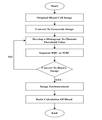
- Original Blood cell images: Original blood cell photos are microscopic images taken with specialist imaging equipment like microscopes and digital cameras. These photos show the biological components of blood, such as red blood cells, white blood cells, and platelets.
- Convert To Grayscale Image: Converting a picture to grayscale entails deleting color information from the original image, resulting in a black-and-white representation with each pixel's intensity proportional to its brightness level. This method is widely used in image processing and computer vision jobs to simplify data while keeping important visual information.
- Develop a Histogram To Obtain Threshold Value: Creating a histogram to determine a threshold value entails examining the distribution of pixel intensities in a picture. The histogram depicts the frequency with which each intensity value appears in grayscale photographs, ranging from 0 (black) to 255 (white). Peaks and valleys on the histogram indicate regions of high and low intensity, respectively.
- Segment WBC Or RBC: Red blood cells (RBCs) and white blood cells (WBCs) are segmented from a microscopic image to allow for further examination. This method is essential in a variety of medical applications, including illness detection and cell counting. One typical method for segmenting RBCs or WBCs is to use image processing techniques like thresholding, which involves applying a threshold value to the image to separate foreground (cells) from background.
- Convert To Binary Image: Converting an image to a binary image entails converting it into a black-and-white representation with each pixel either black (foreground) or white (background). This is often accomplished using thresholding, which applies a threshold value to the grayscale image. Pixels with intensity values higher than the threshold are set to white, while those lower are set to black.
- Image Enchancement: Image enhancement is the process of increasing an image's quality or visual appearance in order to make it better suited for a specific purpose or improve human perception. This can include techniques like changing brightness and contrast, sharpening edges, minimizing noise, and improving colors. Image enhancement seeks to highlight key features, improve clarity, and make images more visually appealing or easy to analyze.
- Ratio Calculation of Blood: Displaying whether a person has a condition or not usually entails displaying diagnostic results or projections based on medical data and analysis. This display of data is critical for informing healthcare decisions and guiding treatment strategies.
VI. ACKNOWLEDGMENT
The group express our gratitude most sincerely to our guide Ms.V.V.Nagamani who guided and motivated us in this course of time of understanding the concepts. We are grateful for the insightful comments offered by the peer reviewers.
Conclusion
In conclusion, we involved in detection the types of leukemia using microscopic blood sample images. We built the system based on typical microscopic images features by observing changes in texture, geometry, colors and statistical analysis as a classifier input. We require our system to be efficient, reliable, less processing time, smaller error, high accuracy, cheaper cost and must be robust towards varieties from individual, sample collection protocols, time and etc. The information extracted from the microscopic images of blood samples will be benefited to the people of being able to predict, solve and treat blood diseases immediately for a particular patient.
References
[1] Diagnosis of Acute Lymphoblastic Leukemia Using Microscopic Blood Cell Images https://www.researchgate.net/publication/361842137_Diagnosis_of_ Acute_Lymphoblastic_Leukemia_Using_Microscopic_Blood_Cell_Images Author:Narendra Mustare [2] Machine Learning in Detection and Classification of Leukemia Using Smear Blood Images: https://www.hindawi.com/journals/sp/2021/9933481/ Author: Farkhondeh Asadi [3] Machine learning in detection and classification of leukemia using C-NMC_Leukemia https://link.springer.com/article/10.1007/s11042-023-15923-8 Author:Samah A. Gamel [4] Automated detection of leukemia by pretrained deep neural networks and transfer learning: A comparison https://www.sciencedirect.com/science/ article/abs/pii/S1350453321001168 Author:K.K. Anilkumar [5] Machine Learning Algorithms For Diagnosis Of Leukemia https://www.researchgate.net/publication/339551705 _Machine_Learning_Algorithms_For_ Diagnosis_Of_Leukemia Author: Devi Thirupathi [6] Purine Metabolism Modulates Leukemia Stem Cell Maintenance in MLL-Rearranged Acute Leukemia https://www.researchgate.net/publicatio n/376163634_Purine_Metabolism_Modulates_Leukemia_Stem_Cell_Maintenance_in_MLL-Rearranged_Acute_Leukemia Author: Lin Tan
Copyright
Copyright © 2024 V. V. Nagamani, Sirigiri Lakshman Sai, Puram Pavan, Chilukuru Venkata Sai. This is an open access article distributed under the Creative Commons Attribution License, which permits unrestricted use, distribution, and reproduction in any medium, provided the original work is properly cited.

Download Paper
Paper Id : IJRASET60399
Publish Date : 2024-04-16
ISSN : 2321-9653
Publisher Name : IJRASET
DOI Link : Click Here
 Submit Paper Online