

Ijraset Journal For Research in Applied Science and Engineering Technology
Review on Diabetic Retinopathy Detection Using Molecular Segmentation In Convolution Neural Network
Authors: Rushikesh Bhusari, Suraj Paswan, Tushar Parate, Sandip Neware, Prabhakar Khandait
DOI Link: https://doi.org/10.22214/ijraset.2022.40122
Certificate: View Certificate
Abstract
People who suffer from diabetes are at risk of developing blindness since their pancreas doesn\'t produce enough insulin. Diabetic retinopathy is a condition in which a patient\'s vision gradually worsens as the disease advances. To better understand how diabetes affects the eye, retinal images captured with a fundal camera can be helpful. This study aims to detect blood vessels, identify hemorrhages, and classify different phases of diabetic retinopathy into normal, mild, and non-proliferative diabetic retinopathy (NPDR). When it comes to classifying the various stages of diabetes-related retinopathy, the retinal picture is used as a starting point. Using the contrast between the blood vessels and the surrounding background, the retinal vascular is segmented.. Density analysis and bounding box approaches were used to identify potential hemorrhage sites. According to blood vessel and hemorrhage area and perimeter, Random Forests technique was used to finally classify various phases of eye disease.
Introduction
I. INTRODUCTION
When the pancreas fails to produce enough insulin or the body is unable to effectively process it, diabetes develops. Chronic damage to blood vessels over time can lead to diabetic retinopathy in people with diabetes. One-tenth of diabetics lose their vision after 15 years, and another two percent suffer from vision damage that is severe. Diabetes affects an estimated 220 million people globally, according to the WHO [1]. It is the sixth leading cause of blindness among Indians over the age of 40, making the country the global epicenter of diabetes.
A fundal camera with a back-mounted digital camera [2] provides useful information regarding the consequences, nature, and status of diabetes's effect on the eye. It is easier for ophthalmologists to organize treatment and monitor progress when they have access to these images [3].The retinal
Unlike other parts of the human circulatory system, microvasculature can be immediately viewed in vivo and imaged for digital image analysis [2].
An enhanced non-parametric approach for identifying retinal hemorrhages is used to classify the retinal cases in this study, which is an improvement over the previously developed matched filter method. Finding blood vessels and identifying hemorrhages are the primary goals of this study. The final goal is to classify the findings into normal, moderate, and severe diabetic retinopathy (NPDR). Retinal illness is a common cause of blindness among the working-age population in western countries, according to international research. This is why it is important to get a diagnosis as soon as possible in order to avoid blindness. A tool for the early diagnosis of diabetic retinopathy and other eye diseases, including macular degeneration associated with ageing, glaucoma, and retinoblastoma, has been developed using fundus photographs. A high-definition laser camera is used to compare photos taken with different color versions inside the eye. These images are known as fundus photos. MATLAB may be used to extract the functions from these fundus images. Automatic screening will help to quickly determine the patient's situation in a more accurate manner. In order to detect diabetic retinopathy-induced macular abnormalities, fundus images can be processed using morphological operations, filters, and thresholds. Glaucoma is caused by an increase in intraocular strain in the affected person's fundus picture. The K-approach clustering set of principles and morphological operation are used to diagnose glaucoma. Retinoblastoma is caused by the growth of a malignant tumor in the retina. Using quick Fourier rework and log transform, the diagnosis of retinoblastoma can be completed Age-related macular degeneration is the primary cause of vision loss in the elderly. Histogram equalization and thresholding are used to diagnose ARMD. The graphical user interface allows for the diagnosis of four retinal diseases using the user's retinal images and their related symptoms. The output of the processing will be shown to the viewer so that they can see how our retina has changed in comparison to the original image.
Diabetic Retinopathy and other retinal diseases (DR). A retinal picture evaluation machine can be built to improve the accuracy of the diagnosis and make their task easier. Diabetic Retinopathy (DR) is the most common eye problem in diabetes. Diabetic retinopathy (DR) is the leading cause of visual impairment and blindness in diabetic people worldwide. For diabetics, early detection and appropriate treatment of diabetic eye diseases can significantly reduce the risk of vision loss. It takes a lot of time and money to review a large number of images with the help of the doctors. DR can lead to a variety of abnormalities in the retina, including micro aneurysms, hemorrhages, hard exudates, and cotton wool patches. Yellow intra retinal deposits of serum lipoproteins are hard exudates. During the formation of exudates, lipid or fat escapes from abnormal blood vessels. If the exudates spread into the macular area, vision loss can result. This research examines the use of Morphological techniques for the detection of exudates in retinal images, and in particular, for the detection of exudates in comparison to ordinary retinal pictures.
We utilize a 3-D mesh if the problem is complex and requires a three-dimensional representation of the continuum. Triangle or quadrilateral shaped elements can be used for area elements. The form and order of the elements is determined by the intricacy of the geometry and the uniqueness of the problem being described. There is no thickness to membrane components. Consequently, they lack bending stiffness and can only carry stresses in the detail plane. The three-dimensional modeling of thin-walled regions is facilitated by the use of plate and shell elements. Assuming the weight is borne by the bend, a spherical plate precept is applied to the plate element. When flexure and membrane movement are combined, shell elements are employed to create a hybrid shell.
First, patients with diabetic retinopathy have their retinas photographed with a digital fundus camera. Red, green, and blue are the primary colors of the original artwork. For further processing, a grayscale version of the original RGB image is created. Afterwards, noise reduction techniques are employed. Use of median filter in this case. Use the median filter to minimize noise while maintaining the sharpness of the image's edges. Using Contrast restricted Adaptive Histogram Equalization, the median filter's output image is improved. The improved image is then subjected to morphological surgery. Morphological operations include dilatation, erosion, closure, and opening.. Closing is used in this instance. The identical intensity values were closed using the closing method. The threshold value is used to turn the morphological result into a binary image. Morphological results will be used to calculate threshold values for grey thresh method. A retinal scan reveals the presence of exudates for the first time
II. OBJECTIVES
With the use of continuous two-dimensional stationary wavelet transform responses at various scales, we were able to construct Feature vectors for use in our project.
Allowing noise filtering and vessel augmentation in one step, the Stationary wavelet is able to turn to specified frequencies.
It is possible to construct complex decision surfaces and compare their performance with the linear lowest squared error classifier by training a neural network with class-conditional probability density functions (likelihoods) characterised as "Gaussian mixtures."
Using morphological processes and segmentation techniques, a reliable system for detecting retinal vessels and exudates from an eye fundus picture will be implemented.
For the detection of retinal vessels and exudates, the combination of a multi-structure morphological approach and a segmentation technique is beneficial. The modules here are. Plane separation, contrast enhancement, and morphological processing are all included in this module for the detection of retinal blood vessels. Detection of exudates in which the Segmentation Method is employed. Out-of-plane distortion is considered to be relevant for plate elements when it is only a few millimeters greater.
III. PROPOSED PLAN OF WORK
- This methodology is effective for achieving histogram equalization that isn't seen in standard methods. Heterogeneous histogram equalization employs the entire image to adjust the brightness, while adaptive histogram equalisation makes use of different areas of the image. As a result, even low-contrast local areas improve in contrast..
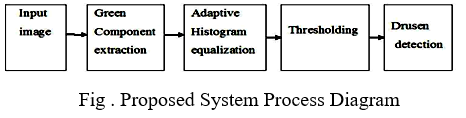
- The term "contrast-limited adaptive histogram equalization" also applies here. For example, the intensity features can be used to identify druse by applying a specific threshold value. The original image (RGB) gets transformed into a grayscale image in retinoblastoma..
- A preprocessing procedure for better outcomes in subsequent processing, median filtering preserves the edges while reducing noise.
- To finish off, a Gaussian filter is applied to the image. For a black and white rendition of the RGB image, the next step is thresholding.
- Fast Fourier Transform is then used to deconstruct an image's real and imaginary components in frequency domains..
- In this image, the frequency is equal to the number of pixels. To extend the black pixels' range into the brilliant zone while plotting the 2D Fourier transform magnitude, we must log transform the pixel values.
- As a result of this, we can more clearly witness the transformation. To acquire images in glaucoma, the source image is retrieved. Correct disc and cup boundaries can be extracted using super pixel segmentation, a computer technique
The single parameter for super pixel segmentation is the number of super pixels. It is the process of dividing an image into a number of distinct parts. In this case, the K-Means clustering algorithm is used..
IV. METHODS
The following points serve as the foundation for classification:
- Extract the retinal blood vessels using Bit Plane separation, Contrast Enhancement, and the Morphological Process. Finally, feature extraction is done using DWT and Energy feature coefficients. Probabilistic neural networks (PNN) are trained on the image.).
- To determine if the retina is normal or pathological, exudates are extracted and classified using segmentation methods like the K-means Clustering approach (algorithm shown below)..
- Morphological operations are applied on segmented image for smoothening the exudates part.
V. METHODOLOGY
The task of accurately detecting the optic disc in color retinal images in an automated retinal image analysis system is significant. In order to distinguish between normal and abnormal retinal characteristics, one must first identify the same. A portion of the optic nerve and blood vessels may be seen extending into the retina from the optic disc . Because of this, it is also referred to as the "blind spot". The diameter of the optic disc varies from patient to patient, but in a normal fundus imaging it is always between 80 and 100 pixels. Image processing, machine learning pattern recognition, and computer visualization all fall under the umbrella of medical image analysis Visual interpretation and analysis of retinal pictures by ophthalmologists is used to identify a wide range of conditions Diabetic Retinopathy and other retinal diseases (DR). A retinal image analysis system can be built to speed up the diagnosing process and make their job easier. In diabetics, Diabetic Retinopathy (DR) is the most prevalent kind of vision loss due to the condition. In diabetics, DR is the leading cause of vision impairment and blindness.
As a diabetic, you should have your eyes checked often to ensure that you aren't at risk of losing your vision. Examining a large number of photos takes a long time and is expensive for doctors. It's not uncommon for DR to induce a variety of retinal abnormalities, such as micro aneurysm and hemorrhage and hard exudates and cotton-wool patches. Serum lipoproteins form yellowish intra retinal deposits known as hard exudates.
Lipid or fat leaks from faulty blood arteries cause exudates to occur. If the exudates reach the macula, vision loss is possible.
Morphological techniques are used in this study to detect exudates in retinal images, and the results are compared to normal retinal images primarily to detect exudates.
VI. WORD SEGMENTATION
- Pixel Segmentation using Super Pixels. In order to determine the disc border, segmentation is applied to the area of interest. A technique known as super-pixel segmentation is employed. It employs a straightforward linear clustering technique. K is the only metric that matters here (the number of super pixels). Accuracy improves as K grows, but assessment time does as well. A shorter computation time is required when K is small, but the results are subpar.
- Clustering is a method of grouping objects into distinct categories. According to the K-means clustering algorithm, all objects in the dataset have a specific placement in three-dimensional space. As close to each other as possible, and as far away from other objects as possible, each cluster is partitioned..
- Morphological Operations It is explained in Diabetic Retinopathy.
- Ratio of Cups to Disks We can figure out the ratio of the diameters of the vertical cups to those of the vertical discs. Vertical Cup Diameter (VCD) and Vertical Disc Diameter (VDD) are both known as CDR. The presence of glaucomatous glaucoma is confirmed when CDR I levels above the threshold.
VII. CLASSIFICATION ALGORITHM
The algorithm is composed of the following steps:
- Initialize U=[uij] matrix, U(0)
- . At k-step: calculate the centers vectors
- C (k) = [cj] with U (k) Cj=(∑uijm*xi)/( ∑uijm)
- Update U (k), U (k+1) uj= [(1) / ( ∑( (||xi-cj||) / (xi-ck) )(2/(m-1)) ]
- If || U (k+1) – U (k)||
Conclusion
A digital image processing strategy combination has been used to develop an automated software application for screening and diagnosing DR. For detecting DR in fundus photos, this software is the most accurate. In remote or rural areas where an ophthalmologist isn\'t available, or if the ophthalmologist is overworked, it can be utilized as an alternate or complementary screening equipment. However, the program\'s classification of DR severity is accurate. To improve the accuracy of DR classification, additional software development is needed, which may include additional virtual picture processing techniques or other methods based on artificial intelligence and deep learning.
References
[1] Pawe? Liskowski, Krzysztof Krawiec, Member, Citation information: DOI 10.1109/TMI.2016.2546227, IEEE Transactions on Medical Imaging “Segmenting Retinal Blood Vessels with Deep Neural Networks”. [2] R. Nekovei and Y. Sun, “Back-propagation network and its configurationfor blood vessel detection in angiograms.” IEEE Transactions on Neural Networks, vol. 6, no. 1, pp. 64–72, 1995. [Online]. Available: http://dblp.uni trier.de/db/journals/ tnn/tnn6.html #NekoveiS95 [3] G. E. Hinton, N. Srivastava, A. Krizhevsky, I. Sutskever, and R. R. Salakhutdinov, “Improving neural networks by preventing co-adaptation of feature detectors,” arXiv preprint arXiv:1207.0580, 2012. [4] Y. Bengio, “Learning deep architectures for ai,” Foundations and trendsR in Machine Learning, vol. 2, no. 1, pp. 1–127, 2009. [5] J. Schmidhuber, “Deep learning in neural networks: An overview,”Neural Networks, vol. 61, pp. 85–117, 2015. [6] A. Krizhevsky and G. Hinton, “Learning multiple layers of features from tiny images,” Computer Science Department, University of Toronto, Tech. Rep, vol. 1, no. 4, p. 7, 2009. [7] A. F. Aqeel and S. Ganesan, “Automated algorithm for retinal image exudates and drusens detection, segmentation, and measurement,” in Proceedings of the IEEE International Conference on Electro/Information Technology (EIT \'14), pp. 206–215, Milwaukee, Wis, USA, June 2014. [8] P. M. Rokade and R. R. Manza, “Automatic detection of hard exudates in retinal images using haar wavelet transform,” International Journal of Application or Innovation in Engineering & Management, vol. 4, no. 5, pp. 402–410, 2015 [9] A. Dosovitskiy, J. T. Springenberg, M. Riedmiller, and T. Brox, “Discriminative unsupervised feature learning with convolutional neural networks,” in Advances in Neural Information Processing Systems, 2014, pp. 766–774. [10] X. Jiang and D. Mojon, “Adaptive local thresholding by verification based multithreshold probing with application to vessel detection in retinal images,” Pattern Analysis and Machine Intelligence, IEEE Transactions on, vol. 25, no. 1, pp. 131–137, 2003 [11] A. Osareh and B. Shadgar, “Automatic blood vessel segmentation incolor images of retina,” Iran. J. Sci. Technol. Trans. B: Engineering, vol. 33, no. B2, pp. 191–206, 2009.
Copyright
Copyright © 2022 Rushikesh Bhusari, Suraj Paswan, Tushar Parate, Sandip Neware, Prabhakar Khandait. This is an open access article distributed under the Creative Commons Attribution License, which permits unrestricted use, distribution, and reproduction in any medium, provided the original work is properly cited.

Download Paper
Paper Id : IJRASET40122
Publish Date : 2022-01-28
ISSN : 2321-9653
Publisher Name : IJRASET
DOI Link : Click Here
 Submit Paper Online