

Ijraset Journal For Research in Applied Science and Engineering Technology
Ensemble Classification Model for Brain Tumor Detection
Authors: Taranjot Kaur, Nikhil Dagwar, Sumit Walke, Pratik Pawar, Anuja Chincholkar
DOI Link: https://doi.org/10.22214/ijraset.2023.55949
Certificate: View Certificate
Abstract
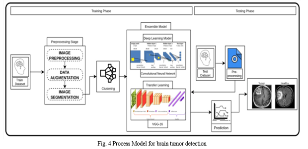
Brain tumor analysis is essential in timely diagnosis and effective treatment to cure patients. The growth of abnormal cells in the brain is the cause of brain tumors, which can lead to cancer. Brain tumors occur due to uncontrolled and rapid cell growth. If not treated at an initial phase, it may lead to death. The variations in tumor location, shape, and size pose a major challenge for brain tumor detection. The aim of this survey is to provide a comprehensive literature on brain tumor detection through magnetic resonance imaging to help the researchers. In this framework based on deep learning, magnetic resonance images (MRIs) are used to identify and categorize brain tumors. In the first step, we propose a preprocessing approach to work only on a small portion of the image rather than the entire image. These little portions of the images provide a very quick and precise brain tumor prognosis, which aids in giving patients the right treatment. The radiologist can make speedy conclusions thanks to these predictions. In the proposed work, A self-defined artificial neural network (ANN) and a network of convolution neural networks (CNN) are used in the proposed work to detect the presence of brain tumors, and their performance is examined.
Introduction
I. INTRODUCTION
The brain is the most important organ in the human body, which controls the entire functionality of other organs and aids in decision-making. The central nervous system (CNS) consists of the brain and spinal cord, which are the primary components of the human nervous system. The tumor is a fibrous mesh of unwanted tissue growth in our brain that proliferates uncontrollably. Brain injury can result from any tumor growing inside the skull, which is a serious risk to the brain. The survival rates for tumors vary depending on their texture, location, and shape. Brain tumors are the tenth most common cause of death. According to the National Brain Tumor Foundation (NBTF), the number of individuals who have died from brain tumors has increased by 300%. The biopsy of brain tumor is not as straightforward as the biopsy of other parts of the body, as it requires surgery. Magnetic Resonance Imaging (MRI) is the most commonly used method for accurate diagnosis without surgery. However, radiologists and clinicians must manually analyse brain MR scans to discover and localise tumours and normal tissues, as well as classify tumours from medical imaging. To address this issue, a computer-aided diagnosis (CADx) system is required. It must be adopted in order to reduce workload and help radiologists or doctors analyse medical images. Several researchers have presented robust and accurate techniques to automate brain tumour identification and classification in the past. A brain tumour is often classified as either primary or secondary tumors. Primary brain tumours form in the brain tissues, whereas secondary tumours form elsewhere in the body and spread to the brain via blood flow. Meningioma, glioma, and pituitary tumours are the most dangerous types of primary brain tumours and the most difficult to identify and treat.
Grades I through IV of a brain tumour are described by the WHO. Tumours of grades I and II are thought to grow slowly, but those of grades III and IV are more aggressive and have a worse prognosis. The specifics of brain tumour grading are as follows in this regard:
Grade I: These tumours develop gradually and do not spread quickly. These can nearly entirely be removed through surgery and are linked to greater long-term survival chances. Such a tumour includes grade 1 pilocyticastrocytoma.
Grade II: Although they develop slowly, these tumours have the potential to invade nearby tissues and progress to higher grade tumours. Even after surgery, these tumours are prone to recurrence. One example of such a tumour is the oligodendroglioma.
Grade III: These tumours can infect the surrounding tissues and grow more quickly than grade II tumours. Such tumours require post-surgical radiotherapy or chemotherapy as surgery alone is ineffective in treating them. Anaplastic astrocytoma is an illustration of one such tumour.
Grade IV: The most aggressive and easily spread tumours are those in question. They might even exploit blood vessels to develop quickly. One such tumour is glioblastoma multiforme.
- Computed Tomography:- Compared to images produced by standard X-rays, computed tomography (CT) images offer more detailed information. Since its introduction, the CT scan has garnered widespread endorsement and adoption. According to a study there are 62 million CT scans performed annually in the USA alone, 4 million of which are on children. The soft tissues, blood arteries, and bones of many human body components are visible on CT scans. More radiation is used than with standard X-rays. Multiple CT scans may expose you to more radiation, which could increase your chance of developing cancer. According to CT radiation exposures, the risks of developing cancer have been calculated. MRI offers strong contrast among the soft tissues, allowing for a sharper view of anatomical structures, and can even assist in evaluating areas that are obscured in a CT scan.
- Magnetic resonance imaging:- An MRI scan is utilised to thoroughly examine various body regions and also aids in the early detection of brain disorders than other imaging modalities. As a result, tumour segmentation is a difficult task due to the complexity of brain structures. The preprocessing strategies, segmentation methods, feature extraction and reduction techniques, classification techniques, and deep learning strategies are all included in this review. The presentation of benchmark datasets and performance metrics concludes.
Recent breakthroughs in machine learning, particularly deep learning, have enabled the recognition and classification of medical imaging patterns. This field has thrived by enabling the retrieval and extraction of knowledge from data rather than depending on specialists or scientific literature. For brain tumor analysis as well these techniques have been used. However, ML-based algorithms need human feature extraction and classification and are only applicable to small amounts of data. Deep learning (DL) has merged feature extraction and classification in a self-learning fashion on a large amount of labelled data, significantly improving performance.
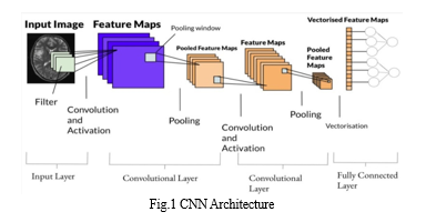
This research paper focuses on the application of CNN (Convolutional Neural Network)-based techniques for brain tumour analysis. However, ML-based algorithms need manual feature extraction and classification and are only applicable to a limited amount of data. Whereas Deep learning (DL) integrated the extraction and classification of features application over large amount of labelled data, significantly improving performance. Furthermore, CNN is a subset of DL that is specifically built for images or two-dimensional (2D) data. It only accepts datasets that have undergone minimal preprocessing and extracts various attributes from MR images without the need for human interaction. Deep CNN models are commonly employed to detect brain tumours. However, brain tumor analysis is highly challenging because of variable morphological structure, tumor appearance in an image, and illumination effects which needs an efficient DL-based brain tumor analysis system to strengthen theradiologist's decision. Convolutional neural network (CNN) convolutional is name of mathematical linear operation. The dimension of the image is reduced at each layer of CNN without the loss of information needed for training. Different processing like convolve, max pooling, dropout, flatten and dense are applied for creating the model.In this research paper we are implementing CNN using transfer learning technique. It is the reuse of a pre-trained model on a new problem.
3. Existing pre-trained CNN Models:- Both the detection and classification models are trained separately in our suggested two-phase structure. Ten well-known customised CNN models are i-VGG-16 , ii-VGG-19 , iii-SqueezeNet , iv-GoogleNet , v-ResNet-18 , vi-ResNet-50 , vii-XceptionNet , vii- 7 InceptionV3, ix-ShuffleNet , and x-DenseNet201 are some of the CNN models that were used. For TR-SC models, all layers of networks changed their weights as a result of our first training of all CNN architectures.
II. EXPERIMENTAL SETUP
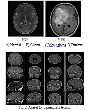
- Data Set:- The MRI brain tumour dataset is used to evaluate the system's performance. The publicly accessible database, Kaggle, served as the source of the dataset. Kaggle enables users to discover and share various datasets, collaborate with various machine learning and deep learning publishers and data scientists, and develop and experiment with various models in various data science environments. This dataset is further divided into two parts i.e images with the tumor and images without the tumor wherein the images with tumor are further classified into three types of tumors- Glioma, Pituitary, Meningioma.
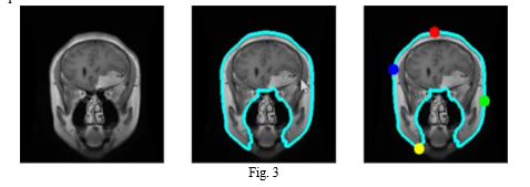
2. Segmentation:- Segmentation takes the needed area out of the input photos.Therefore, accurately segmenting lesion sites is a more important task. Semi- and completely automated approaches are used since the manual segmentation procedure is flawed. Semi-automated segmentation techniques outperform manual segmentation in terms of results. The three types of semi-automated methods are initiation, evaluation, and feedback response. Segmentation from input photos, segmentation extracts the necessary region.Therefore, accurately segmenting lesion sites is a more important task. Semi- and completely automated approaches are used since the manual segmentation procedure is flawed. When compared to hand segmentation, semiautomated approaches produce results that are acceptable. The three types of semi-automated methods are initiation, evaluation, and feedback response.
3. Thresholding:- The thresholding method is a straightforward and effective way to segment the necessary objects , but choosing an optimised threshold in low-contrast images can be challenging. Based on image intensity, threshold values are chosen using histogram analysis. There are two types of thresholding techniques: local and global. The global thresholding method is the best choice for segmentation if the objects and backdrop have highly homogenous contrast or intensity. The Gaussian distribution approach can be used to find the ideal threshold value. These techniques are used when the threshold value cannot be determined from the entire image histogram or when a single threshold value does not produce satisfactory segmentation results.
4. Noise Reduction:- Noise reduction is a common task in digital image processing, where you try to remove unwanted or random variations in pixel values from an image. Noise can degrade the quality and clarity of an image, and affect its usefulness for analysis or display.
5. Feature Extraction :- The features are extracted from the input training data using convolutional layers. There are a number of filters built into each convolution layer that aid in feature extraction. In general, the complexity of the features learned by convolution layers grows as the depth of the CNN model decreases. For instance, the initial convolution layer of training samples collects simple features whereas the last convolution layer captures complicated features. According to the CNN model's design, this process of sequentially transferring input through the convolution and pooling layers is repeated. This method is performed 2-4 times for educational purposes.
6. Training Model:- The output from a multi-layer neural network is then applied to the output from subsequent convolution and pooling layers. Here, each unit of a neuron serves as a feature map that contains data about a unit. On a training dataset with these feature maps, the model is trained. It consists of sets of relevant input data that affect the output as well as sample output data. After this the model is evaluated and validated on an unseen data i.e testing data.
Conclusion
Globally greater mortality rates can be significantly reduced by early identification of brain tumours. The accurate detection of brain tumours is still quite difficult because of the tumor\'s shape, shifting size, and structure. The classification of MR images has a significant impact on the clinical diagnostic and therapy choices made for patients with brain tumours.With the help of several MR scans, we present a CNN model for the early diagnosis of brain tumours and saw encouraging results. One of the greatest methods for analysing the image collection is CNN. CNN performs the prediction by cropping the image to the desired size while preserving the necessary data. This model was created using the trial and error methodology.In our strategy, such patches are chosen to inform the network that the tumor\'s centre is situated inside this area after extracting the tumor\'s predicted area utilising a potent preprocessing methodology wherein the tumor can also be further classified.
References
[1] Park JG, Lee C (2009) Skull stripping based on region growing for magnetic resonance brain images. Neuroimage 47:1394–1407 [2] Litjens, G., et al., A survey on deep learning in medical image analysis. Medical image analysis,2017 [3] Somasundaram K, Kalaiselvi T (2010) Fully automatic brain extraction algorithm for axial T2-weighted magnetic resonance images. Comput Biol Med 40:811–822 [4] Roy S, Bandyopadhyay SK (2012) Detection and quantification of brain tumor from MRI of brain and it’s symmetric analysis. Int J Inf Commun Technol Res 2:1–7 [5] Srinivas B, Rao GS (2019) Performance evaluation of fuzzy C means segmentation and support vector machine classification for MRI brain tumor. In: Soft computing for problem solving. Springer, New York, pp 355–367 [6] Agn M, Puonti O, af Rosenschöld PM, Law I, Van Leemput K(2015) Brain tumor segmentation using a generative model with an RBM prior on tumor shape. In: BrainLes vol 2015, pp 168–180
Copyright
Copyright © 2023 Taranjot Kaur, Nikhil Dagwar, Sumit Walke, Pratik Pawar, Anuja Chincholkar. This is an open access article distributed under the Creative Commons Attribution License, which permits unrestricted use, distribution, and reproduction in any medium, provided the original work is properly cited.

Download Paper
Paper Id : IJRASET55949
Publish Date : 2023-09-30
ISSN : 2321-9653
Publisher Name : IJRASET
DOI Link : Click Here
 Submit Paper Online