

Ijraset Journal For Research in Applied Science and Engineering Technology
Framework for Brain Disease Detection Using Machine Learning Approach
Authors: Mir Kaisar Bashir, Talwinder Kaur
DOI Link: https://doi.org/10.22214/ijraset.2022.44436
Certificate: View Certificate
Abstract
In healthy persons, machine learning analysis of neuroimaging data can reliably predict chronological size/age, and departures from healthy brain ageing are linked to cognitive impairment and illness. Most brain illnesses, such as epilepsy or a brain tumor, are difficult to diagnose and need several visits to doctors and electroencephalogram technicians. Using Artificial Intelligence and deep learning, this research attempts to automate brain disease detection based on brain size. Many brain diseases can be identified by measuring the size of the brain. Using a noninvasive method to collect data directly from the brain provides substantial information about its health and illness. When examining an Electroencephalography, clinicians presently classify and discover abnormalities on the brain. It may be feasible to learn and categories brain activity with the proper quantity of data and machine learning algorithms (i.e.: anxiety, epilepsy spikes, abnormal tumor activity, disease etc.). Following that, hybrid approach would analyses the brain datasets and look for signs of a condition in order to automate the identification and categorization of the disorders/diseases discovered.
Introduction
I. NTRODUCTION
The last three centuries in a huge percentage of un-structuring data was generated for hospitals and healthcare systems in connection with such data (MIDs), genomics as well as free text and data flux from the tracking instruments. The study of evidence considerably altered the methods used by medical professionals and physicians on the diagnosis, interpretation and recovery of brain diseases, risk recognition for, and exposure to, interventions are involved.
In reality, IL and PM therapy started a progressive movement in the area, laying out a very clear and noninvasive testing, evaluation, treatment, management, and diagnosis in terms of disease conditioning. Costly image recognition systems have benefited from the discoveries in a more cost-effective and low-risk framework.
For eg, a positron emissions tomography (PET) camera and magnetic resonance imaging (MRI) to enhance brain thought by allowing doctors to perform non-invasive brain anatomy experiments and predict the reasons for pathological events, to adjudicate pathology linked to different diseases, Circularly Measured Tomography (CT).
II, LITERATURE WORK
However, according to Cestniket al., (1987), the transaction must imply that the resulting design was very accurate, low transparency or high transparency, but low performing in terms of general diagnostics.
In Kononenko, (2001) Because of the high level of transparency awareness, empirical research have shown that physicians prefer diagnoses and details of Bayesian classifiers and categories of option tree like Assistant-R and LFC.
In Shattuck and Leahy, (2002) It is critical to adhere to all current criteria in connection to various ways to disease digital administration when it comes to the consequences for health diagnostic determinations. Machine learning algorithms are becoming increasingly useful for simpler tasks such as medical division and estimate.
In Krizhevsky et al (2012) On the other hand, high-efficiency ML algorithms that use artificial neural network algorithms to investigate issues like imagery breakdown on photographs of brain cells or even the much simpler function of comprehension and plausible items have low skills that have straightforward coverage and the abilities of clarification in natural scenes.
III. PROBLEM FORMULATION
Inability to commit absence of brain type II anomaly in brain tissue is not enough, but
Low false alarm incorrect with Type I mistake is also still needed because it aims to help the diagnostic service process and not to substitute individual radiology Assistants for their requirements. A highly educated, talented radiologist has taken several hours to guarantee the diagnostic technique of a specific situation with permission to try out a new way by bringing the associated price advantages.
A. Objectives
- To study and analyze the various approaches followed in Machine Learning using different classifiers to design the diseases detection system.
- To Predication of Diseases on the basis of size of brain.
- To validate the purposed detection system on the basis of different parameter using ensemble hybrid approach.
IV. WORK DONE
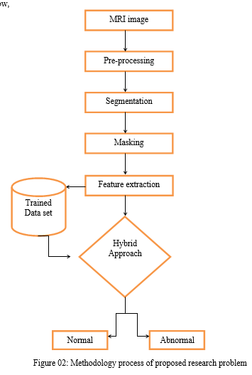
A. Methodology
The process flow is follow,
B. Historical Data
Gathering historical data is the most critical step in solving a machine learning challenge. Historical data in the form of photographs has been entered into the research topic to explain the quantity and quality of data. Our model's accuracy is determined by the quantity and quality of data available.
- Data Formatting: This step is required to remove noise from the data input. We remove noise from the data before giving it to the algorithm to be trained. Organizing any data that might be needed (correct errors; remove duplicates, normalization, deal with missing values, and conversions of data types, etc.).
- Define Parameters: We must partition the data into independent and dependent variables after eliminating the noise from the photos in order to train the model by passing the input data as independent variables and specifying the parameters.
- Training Process and Trained Model: Using data from a sample dataset, we trained an 80 percent dataset and then used that data to feed an 80 percent dataset for testing. We utilized 20 percent of the dataset for testing and observed the results with some standard assumptions that we evaluated and predicted that the model would operate.
- Forecasting Process: On a brain image dataset of about 400 samples, we used CNN, NB, and the proposed model to make the prediction. In order to determine the model's exact efficiency, we'll combine dataset value with time duration.
Conclusion
But there is a dedication to the application of ML in the process of neuroimaging and that could well lead to substantial achievements in the quest for biomarkers used to diagnosis diseases through pictures while still at an early stage. Nevertheless, before ML hits its optimum of neuroimaging ability, a sequence of transformations is expected. Second, in view of the complexities of ML models, we need to step away from experiments of limited sample sizes to moderate sizes in favour of basic images. The effectiveness of this is accomplished by a series of data centres operating together and using the same employability and evaluation processes on various locations in terms of data processing. A second approach to maximize the sample size is through multi-site data sharing initiatives. And second, convergence from CNN and repetitive neural systems is expected to produce major ML improvements over the coming years. During the neuroimaging, the mixture has become especially helpful when a study of FMRI data processes. The ability of ML models, in the end, to learn detailed and uncommon representations through nonlinear changes makes this a positive technique regarding uniform imaging theme foretelling. Though significant issues need to be addressed now, the result discussed here offers early evidence supporting the possible role that ML plays.
References
[1] Alexander, D. C., Dyrby, T. B., Nilsson, M., and Zhang, H. (2017). Imaging brain microstructure with diffusion MRI: practicality and applications. NMR Biomed. doi: 10.1002/nbm.3841. [2] Alpaydin, E. (2014). Introduction to Machine Learning. Cambridge, MA: MIT Press. Altaf, T., Anwar, S. M., Gul, N., Majeed, M. N., and Majid, M. (2018). Multiclass Alzheimer’s disease classification using image and clinical features. Biomed. Signal Proc. Control 43, 64–74. doi: 10.1016/j.bspc.2018.02.019 [3] Amato, F., López, A., Pena Mendez, E. M., Vanhara, P., Hampl, A., and Havel, ? J. (2013). Artificial neural networks in medical diagnosis. J. Appl. Biomed. 11, 47–58. doi: 10.2478/v10136-012-0031-x [4] Ardekani, B. A., Guckemus, S., Bachman, A., Hoptman, M. J., Wojtaszek, M., and Nierenberg, J. (2005). Quantitative comparison of algorithms for inter-subject registration of 3d volumetric brain MRI scans. J. Neurosci. Methods 142, 67–76. doi: 10.1016/j.jneumeth.2004.07.014 [5] Bakas, S., Akbari, H., Sotiras, A., Bilello, M., Rozycki, M., Kirby, J., et al. (2017a). Segmentation labels and radiomic features for the preoperative scans of the TCGA-LGG collection. Cancer Imaging Arch. 286. doi: 10.7937/K9/TCIA.2017.KLXWJJ1Q. [6] Bakas, S., Akbari, H., Sotiras, A., Bilello, M., Rozycki, M., Kirby, J. S., et al. (2017b). Advancing the cancer genome atlas glioma MRI collections with expert segmentation labels and radiomic features. Sci. Data 4:sdata2017117. doi: 10.1038/sdata.2017.117 [7] Belaroussi, B., Milles, J., Carme, S., Zhu, Y. M., and Benoit-Cattin, H. (2006). Intensity non-uniformity correction in MRI: existing methods and their validation. Med. Image Anal. 10, 234–246. doi: 10.1016/j.media.2005.09.004 [8] Bengio, Y., Courville, A., and Vincent, P. (2013). Representation learning: a review and new perspectives. IEEE Trans. Pattern Anal. Mach. Intell. 35, 1798–1828. doi: 10.1109/TPAMI.2013.50 [9] Beni, L. H., Mostafavi, M. A., Pouliot, J., and Gavrilova, M. (2011). Toward 3d spatial dynamic field simulation within GIs using kinetic voronoi diagram and Delaunay tetrahedralization.
Copyright
Copyright © 2022 Mir Kaisar Bashir, Talwinder Kaur. This is an open access article distributed under the Creative Commons Attribution License, which permits unrestricted use, distribution, and reproduction in any medium, provided the original work is properly cited.

Download Paper
Paper Id : IJRASET44436
Publish Date : 2022-06-17
ISSN : 2321-9653
Publisher Name : IJRASET
DOI Link : Click Here
 Submit Paper Online