

Ijraset Journal For Research in Applied Science and Engineering Technology
Green Synthesis, Characterization and Antibacterial Activity of Copper Oxide Nanoparticles Using Annona muricata Stem Extract
Authors: Shivani Kushwaha, Dr. Poonam Prakash
DOI Link: https://doi.org/10.22214/ijraset.2021.39498
Certificate: View Certificate
Abstract
Nanotechnology is a rising field of science and technology that deals with the particles having size in the range of 1 to 100 nm. Copper oxide nanoparticles has many properties like antifungal activity, antibacterial activity, optical properties, conductive properties, etc. Due to its demand of diversified use, copper oxide nanoparticles were fabricated using ecofriendly and non-toxic Annona muricata stem extract. The extract with copper sulphate pentahydrate showed gradual change in the colour of the extract from brown to green which indicates the CuO nanoparticles synthesis. The fabrication is followed by characterization of CuO nanoparticles using UV-vis spectroscope, FTIR, XRD and SEM. The characterization showed roughly spherical shaped nanoparticles in the range of 100nm with high crystalline monoclinic phase. FTIR absorption spectra conclude that the compounds attached with copper oxide nanoparticles could be polyphenols with aromatic ring. The CuO nanoparticles exhibited antibacterial activity; it showed the maximum activity against E.coli (18 mm).
Introduction
I. INTRODUCTION
Green nanoscience or nanotechnology utilizes the application of green chemistry principles for designing of nanoscale products, development of nanomaterial production methods and the applications of nanomaterials.
Since the dawn of medicines, natural products, especially those derived from plants have helped mankind to sustain its health. Over the past century, the phytochemicals in plants have been a pivotal pipeline for pharmaceutical discovery. The importance of the active ingredients of plants in agriculture and medicine has stimulated significant scientific interest in the biological activities of these substances [11].
Despite these studies only a restricted range of plant species has experienced detailed scientific inspection but the knowledge is comparatively insufficient concerning their potential role in nature. Hence, the attainment of a reasonable perception of natural products necessitates comprehensive investigations on the biological activities of these plants and their key phytochemicals.
In a pharmaceutical landscape, plants with a long history of use in ethno medicine are a rich source of active phytoconstituents that provide medicinal or health benefits against various ailment and diseases.
Annonaceae family is the largest family of magnolia order with 129 genera and about 2,120 species. The family consists of tree, shrubs, and woody climbers found mainly in the tropics, although a few species extend into temperate regions. Many species are valuable for their large pulpy fruits, some are useful for their timber, and others are prized as ornamentals.
Annona muricata Linn. is a lowland tropical fruit-bearing tree in the Annonaceae family. Annona muricata is also commonly known as Graviola or Soursop or Gunbanana. The name soursop is due to sour and sweet flavour of its large fruit. Related species includes cherimoya (Annona cherimola) and sugar-apple (Annona squamosa); paw paw (Asimina triloba) are also in the family. The soursop is native to tropical Central and South America and the Caribbean, but is now widely cultivated in tropical areas worldwide, including southern Florida and Southeast Asia, from sea level to altitudes of around 1150 meters.
Soursop is one of most commonly used medicinal plants in Caribbean. Pulp of the fruit is eaten and used as an ingredient in many foods and beverages. Tea is drunk daily and often mixed with other herbal decoctions.
Annona muricata has a wide potent anticancerous agents coined as Acetogenins which play a key role towards many varieties of cancer. Acetogenins are potent inhibitors of NADH oxidase (nicotinamide adenine dinucleotide phosphate-oxidase) of the plasma membranes of cancer cells. The fruit is of economic value and hence cultivated and used widely as an edible food. The plant possess the major pharmacological activities includes cytotoxic, antileishmanial, wound healing, antimicrobial activity. It also has the anticarcinogenic and genotoxic effect. Phytochemical analysis of the plant revealed the presence of tannins, steroids and cardiac glycosides which are the major phytochemical compounds [3].
There is an approach for the synthesis of stable and monodispersed silver nanoparticles at ambient/low temperature using Allium Sativum (garlic) extract as a silver salt reducing agent as well as the postsynthesis stabilizing ligands[4].
Antibiotics become less effective on microorganisms due to many reasons like microbial drug target proteins are changed, inactivation of drug or enzymatic degradation, decrease of membrane permeability, increased efflux of drugs, etc. [7]. Hence, there is an increased need for finding ways to make antibiotics by a different process.
These findings strongly suggest the potential use of Annona muricata as a natural source of antioxidants that can be used as a reducing agents to form copper oxide nanoparticles. Biosynthesis of copper oxide nanoparticles and using it for antimicrobial activity is an important area of research in bio-nanotechnology.
This is an emerging ecofriendly science of synthesizing nanoparticles of well defined, shape, size and controlled monodispersity. The recent studies researcher proposed by a green process for the production of copper oxide nanoparticles. Copper nanoparticles due to their unique biological, chemical and physical properties and the low cost of preparation have been of great interest. Copper nanoparticles can easily oxidize to form copper oxide.
II. MATERIALS AND METHODS
A. Materials Required
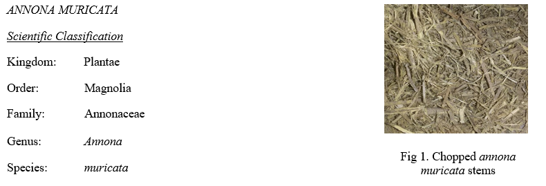
Materials, chemicals and bacterial strains required for experiment were: Air dried finely chopped Annona muricata stems, analytical grade copper sulphate pentahydrate, Mueller Hinton Broth (TM media) and Agar-agar of bacteriological grade (Fisher Scientific) for the media preparation, Oxytetracycline (10,000 ppm) as a positive control and distilled water.
Escherichia coli and Staphylococcus aureus strains as a gram- negative and gram-positive bacteria respectively.
B. Preparation of Extract
Fresh Annona Muricata stems were collected from the umme orgo fruits (agency), Bangalore. The stems were washed and dried for 15-20 days under shady condition and then they were finely chopped as small as possible.
The finely chopped stems were weighed 10g and mixed in 100ml of distilled water for the preparation of extract. This mixture was boiled at 800 C for 30 minutes. The above prepared extract was cooled at room temperature and filtered using Whatmann No.1 filter paper making sure no solid particles were in the filtrate. The filtrate was stored in a conical flask tightly seal packed with aluminium foil and kept at room temperature for further use [10].
C. Preparation of 1mM stock Solution of Copper Sulphate (CuSO4.5H2O)
Analytical grade Copper sulphate solutions was used for the preparation of desired nanoparticles. A 1mM stock solution of CuSO4.5H2O was prepared in distilled water. For this 0.2496 g of copper sulphate was taken in a beaker and 100 ml of distilled water was added dropwise along with the swirling to dissolve the salt homogenously. The beaker containing this solution was tightly covered with aluminium foil and kept away from light.
D. Synthesis of Plant Mediated Copper Oxide Nanoparticles
For the reaction mixture,50ml of the prepared copper sulphate solution was taken from the former prepared 100ml copper sulphate solution in a clean and dry beaker and placed it on the magnetic stirrer at a temperature of 40-50o C. 10ml of the prepared plant extract was added to the beaker. With the addition of extract the colour of the copper salt solution changes from blue to bright green. It was kept stirred for 2 hours, further the mixture was placed in a preheated furnace at 400o C for 5 minutes.
E. Purification of Prepared Nanoparticles
The reaction mixture was centrifuged at the speed of 8000 rpm and the supernatant was taken out by aspiration. The residue was washed with double distilled water thrice and centrifugation was repeated at 8000 rpm for 30 minutes further the powder was collected and dried in hot air oven at 110o C. These oven dried particles were used for further analysis for size, shape, chemical composition and also for performing antibacterial activity.
F. Characterization of Copper Oxide Nanoparticles
The phytofabricated copper oxide nanoparticles were characterized by different methods. The initial formation of the copper oxide nanoparticles was observed by the colour change from blue to sea green which was further confirmed by the following techniques.
- UV-Visible Spectrophotometer analysis: The synthesized copper oxide nanoparticles were characterized by Double Beam UV-Vis spectrophotometer: 2202 by continuous scanning from 200-800nm at Cytogene Research and Development (Lucknow). The distilled water was used as blank.
- Fourier transform infra-red (FTIR) analysis: PerkinElmer spectrum FTIR spectrometer was used for studying the chemical composition of the synthesized nanoparticles. A Fourier transform Infra-Red (FTIR) spectrometer was used to obtain the infra-red spectra of the absorbance and emission of copper oxide nanoparticles. The synthesized sample was determined by OMNIC software. The dried powder was scanned in the range of 4000-500 cm-1 at Department of Chemistry, University of Allahabad.
- X-Ray Diffraction (XRD analysis): The grain size and the phase variety of the synthesized copper oxide nanoparticles was determined by X-ray diffraction spectroscopy. Cu-Kα radiation was used to study the synthesized copper oxide nanoparticles at a voltage of 30 kV and current of 20 mA with scanning rate 0.030/s. Different phases present in the synthesized samples were determined by X’Pert high score software. The crystallite size of the prepared samples was calculated by Debye-Scherrer’s equation
- Scanning Electron Microscopy (SEM) analysis: The surface morphology of the biologically synthesized copper oxide nanoparticles was analyzed using SEM. The sample was examined using electron probe microanalyzer, model no. JXA 8100, JEOL, Japan at an accelerating voltage of 10 keV and current of 1nAmp. Before analysis sample was coated with 20nm thick layer of graphite using vacuum evaporator.
G. Antibacterial activity
- Test organism for antibacterial activity: For determining the antibacterial activity one-gram positive and one-gram negative bacterial strains were used. The bacterial pathogens were collected from the Cytogene Research and Development, Lucknow. Bacterial strains used are listed in the table below:
Table 1. List of bacteria
|
S. No. |
Name of the bacteria |
Bacterial type |
|
1 |
Escherichia coli |
Gram negative |
|
2. |
Staphylococcus Aureus |
Gram positive |
The bacterial strains given in the above table were grown and maintained on a nutrient agar slant at 370 C followed by incubation for 5 days. The culture was stored at 40C for further use. The organisms were sub-cultured once in every 15 days.
2. Media and colloidal solution of nanoparticles preparation: In this study, Mueller Hinton Agar was used which supports growth of a wide range of bacteria including Escherichia coli and Staphylococcus aureus. For preparing Mueller Agar medium, 2.625g Mueller Hinton Broth and 1.875 g of Agar-Agar were added to 125ml distilled water in a 300ml conical flask. This cotton plugged conical flask was sterilized in an autoclave at 1210C for 15 mins. It was then distributed equally (about 25 ml each), inside a laminar hood, on petri dishes while it was still hot (450C) making sure no bubble formation takes place and was allowed to solidify. For preparing the colloidal solution of nanoparticles 50μg and 100μg of nanoparticles were dispersed in 1 ml distilled water.
3. Method of testing the antibacterial activity of the synthesized copper oxide nanoparticles: Antibacterial activity of the synthesized copper oxide nanoparticles was carried out by Agar Well Diffusion Method [14],[18] against Gram negative (Escherichia Coli) and Gram positive (Staphylococcus aureus) bacteria. The pure culture of organism was sub-cultured in nutrient broth. The Mueller Hinton Agar plates were prepared by transferring the hot autoclaved molten media into sterile petri-plates. For bacterial growth, a culture was prepared by spreading the 60μl of the bacterial strains on each MHA plate with the help of a sterile glass spreader. Plates were left standing for 10 mins to let the culture get absorbed. Then about 6 mm (size) wells (4 wells) were punched at four corners of each petri-plates at 1.5 cm away from the disc walls into the MHA plates for testing the antibacterial activity. Using a micro-pipette 20μl each of 50μg/ml, 100μg/ml, positive control (oxytetracycline) and negative control (copper sulphate solution) was poured in each of the wells respectively [26]. After adding the samples in the wells, the petri-plates were kept in a refrigerator for an hour for the absorption of the sample into the surrounding medium from the well. Then the plates were transferred into an incubator set at 370 C to allow the bacterial growth on the medium. After 24 hrs the plates were taken out of the incubator and observed for the zone of bacterial growth inhibition around the wells. The zone of inhibition was measured in millimeter.
III. RESULTS AND DISCUSSION
A. Synthesis of Copper Oxide Nanoparticles
In this study, natural synthesis of CuO NPs was successfully accomplished by using Annona muricata stem extract as it was known to contain abundance of bioactive compounds such as polyphenols, alkaloids, megastigmanes, flavonol triglycosides, phenolics, cyclopeptides and essential oils [13]. The initial identification of the synthesized copper oxide nanoparticle using Annona muricata stem extract was done by the visual change of the colour of solution from blue to bright green which indicates the presence of copper oxide nanoparticles.[17],[22].
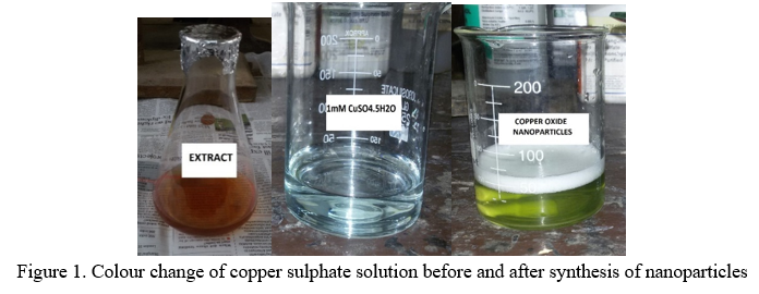
The Annona muricata stem extract does not show any colour change even if it is kept for several weeks until the copper sulphate solution was added to it. Hence the colour of the solution became light green due to the surface plasmon resonance of the deposited copper oxide nanoparticles.
Researcher have reported that the synthesis of CuO nanoparticles can be monitored by the formation of SPR between 250-300nm. However, these differences may be attributed to the difference in the methods used (chemical or biological).
B. Characterization of prepared copper oxide nanoparticles
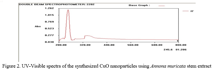
- UV-Visible Spectrum analysis: The UV-Visible spectroscopy is an important technique to determine the formation and stability of the synthesized nanoparticles. The UV-Visible spectroscopy recorded from the nanoparticle solution showed a characteristic surface plasmon resonance (SPR) with absorbance at 200-800 nm range. The peak at 245.6nm is attributed to the formation of CuO nanoparticles and another absorbance peak observed at 335nm indicated the optical band gap in CuO NPs (Fig.2). The result is in close accordance with the result obtained by Prasad et al.,2017. The peak showed that the particles were monodispersed. The exact position of the SPR band may shift, depending upon the properties of the individual particle including size, shape and capping agents [2]. Other researchers have recorded SPR spectra with the maximum absorbances around 220nm [21]. Another similar results was at 350nm along with a small absorbance around 231nm [12].
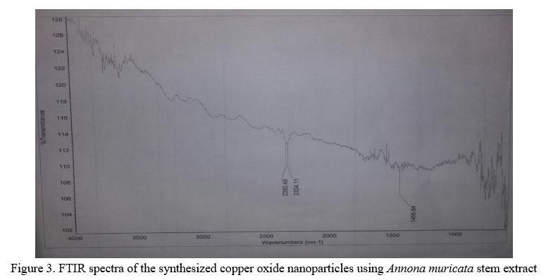
2. Fourier Transform Infra-red (FTIR) Spectrum Analysis: FTIR characterization is used to find the molecules and functional groups present in the sample. After the synthesis of copper oxide nanoparticles, the spectrum showed a number of peak in the region 500-800cm-1 along with peak at 530 which is attributed to vibrations of Cu-O, confirming the formation of nanoparticles (Fig.3). The band at 2350 shows the presence of C=O [8]. The FTIR spectra showed the presence of different functional groups like alcohols(-OH) . The region between 2700 and 3750 cm−1 is known as the OH-stretching region. Peaks reported at 533 cm−1 [9] and 529 cm−1 [8] in the FTIR spectra for CuO-NPs which nearly matches with our results. Therefore, the metal-oxygen frequencies observed for CuO-NPs are in close agreement with that of literature values. Other peaks responsible for the capping and stabilization of nanoparticles with various phytoconstituents and functional groups responsible for these peaks are listed in the Table.2 below:
Table 2. List of peaks and corresponding functional groups
|
Peak value(cm-1) |
Functional group responsible |
Reference |
|
3329,3313 |
O-H stretching of phenol |
[26] |
|
3471,3537 |
N-H stretching of amide |
[22] |
|
3453 |
O-H stretching of H-bonded alcohol |
[17] |
|
2362-2374 |
COO- stretching |
[5] |
|
1635,2350 |
C=O stretching of ketone/ acid present in residual plant material |
[16],[8] |
|
1342 |
C-H bending vibration due to alkanes |
[19] |
|
900-700 |
C-H group bending vibration |
[19] |
|
575,549,561,573,525,584 |
Cu-O stretching |
[15],[5] |
3. X- ray diffraction analysis: Figure. 4 shows the XRD pattern of synthesized copper oxide nanoparticles. All the peaks of CuO NPs can be indexed to the monoclinic crystal system CuO. It showed distinct diffraction peaks at 2θ=32.08o, 35.50, 39.2o, 46.9o, 53o, 68.7o which matches the values of monoclinic-phase CuO reported in the literature (JCPDS card no. 45-0937) [1] suggesting high quality of crystalline CuO prepared. Very few characteristic impurity peaks were also detected which may be due to possible Cu2O impurity. The sharp Bragg’s peaks may be due to capping agent stabilizing the nanoparticles.. The broadening of the peaks in the XRD pattern of the solids is generally attributed to particle size effects. Broader peaks signify smaller particle size and reflects the effects due to experimental conditions on the nucleation and growth of the crystal nuclei. The data are also in very close accordance with the values of monoclinic CuO provided by ICSD (Inorganic Crystal Structure Database, Reference code:01-089-5897) collection code-87124. The comparison is given in the Table 3.
The crystallite size has been estimated from the XRD pattern using the Scherrer’s Equation [5]. The size of the crystallite was in the range of 31-146 nm, which indicated high surface area to volume ratio. Crystal size obtained due to various peak position along with their full width at half maximum (FWHM) is summarized below in the Table 4.
Table 3. Comparision of CuO NPs and ICSD data
|
EXPERIMENTAL NANOPARTICLE (COPPER OXIDE) |
ICSD (Reference code:01-089-5897) Collection code-87124 |
||
|
2θ of peaks |
d- spacing (?) |
2θ of peaks |
d-spacing (?) |
|
32.0872 |
2.78 |
32.526 |
2.75 |
|
35.5720 |
2.52 |
35.468 |
2.52 |
|
39.2033 |
2.36 |
38.969 |
2.32 |
|
46.9688 |
1.93 |
46.304 |
1.95 |
|
53.0328 |
1.72 |
53.480 |
1.71 |
|
56.1521 |
1.63 |
56.722 |
1.62 |
|
68.7860 |
2.32 |
68.125 |
1.37 |
|
67.4710 |
1.38 |
67.928 |
1.37 |
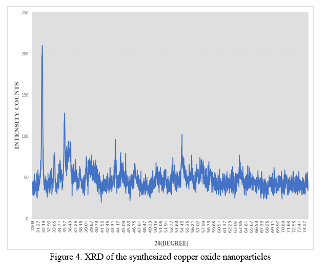
Table 4. Crystallite size corresponding to various peaks
|
Peak position (2θ) |
Peak width/FWHM (2θ) |
Crystal size(nm) |
|
31.9723 |
0.0960 |
89.94 |
|
32.0872 |
0.0590 |
146.39 |
|
34.0072 |
0.1968 |
44.11 |
|
35.5720 |
0.0787 |
110.77 |
|
39.2033 |
0.1574 |
55.98 |
|
43.0080 |
0.811 |
75.55 |
|
46.9688 |
0.1968 |
45.99 |
|
53.0328 |
0.1574 |
58.94 |
|
54.5417 |
0.1574 |
59.34 |
|
63.9163 |
0.3149 |
31.07 |
|
67.4710 |
0.0720 |
138.65 |
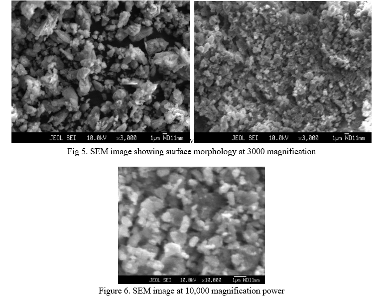
4. Scanning Electron Microscope (SEM) analysis: The morphological studies of synthesized copper oxide nanoparticles were analyzed by SEM. The SEM study demonstrated that the average size of the particles was 100nm as shown in the Fig. 5 at magnification power of 3000 and and Fig.6 with 10,000 magnification power. These figures suggested the aggregation of particles with small grains. Most of the CuO NPs were aggregated with irregular spherical shape. It may be due to the protein which were bound on the surface of the nanoparticles. Such variations in shape and size of the nanoparticles synthesized by biological system is common. Large nanoparticles were seen due to aggregation. The electrostatic interactions and hydrogen bond between bio-organic capping molecules are responsible for the synthesis of copper oxide nanoparticles using plant extract (Thamer et al., 2018). Almost roughly spherical shaped nanoparticles were formed with an average size of 100 nm.
5. Antibacterial activity of copper oxide nanoparticles: Fig 7 and fig.8 shows that synthesized NPs exhibit the zone of inhibition of CuO nanoparticles at different concentration against a gram-negative bacterium (Escherichia coli) and gram-positive bacteria (Staphylococcus aureus). They clearly indicate that the copper oxide nanoparticles inhibit the growth of both gram-negative and positive bacteria and the zone of inhibition increases with the increase in concentration of nanoparticles which are shown in Table 5. It is clear from Table. 5 that CuO nanoparticles have shown almost similar antibacterial activities against both the pathogens. The variation in the sensitivity or resistance to both gram-positive and gram-negative bacteria populations could be due to the differences in the cell structure, physiology, metabolism or degree of contact of organisms with nanoparticles.
Table 5. Zone inhibition of copper oxide nanoparticles against bacterial strains tested
|
|
Zone of inhibition in (mm) |
||||
|
S NO. |
Bacterial strain |
Positive control(20μl) (oxytetracycline) |
Negative control (copper sulphate 100μg/ml) |
CuO NPs (50μg/ml) |
CuO NPs (100μg/ml) |
|
1. |
Escherichia coli |
22±1 |
- |
14±1 |
18±1 |
|
2. |
Staphylococcus aureus |
27±1 |
- |
11±1 |
16±1 |
These results demonstrate the excellent antibacterial behavior of CuO nanoparticles synthesized at low temperature. Broadly, interactions between the negative charges of microorganisms and the positive charges of nanoparticles produce an electromagnetic attraction between the microbe, and effective levels of active nanoparticles. Such interactions lead to oxidation of surface molecules of microbes resulting in their death. Bio destructive effects such as degradation of deoxyribonucleic acid were observed following exposure of gram-positive bacteria to silver and copper nanoparticles by other works [24].
The antibacterial mechanism of copper oxide nanoparticles has been attributed to the fact that Cu+ ions eluted from nanoparticles are absorbed by bacteria when the nanoparticles concentration is high enough. Copper ions are absorbed onto the bacterial cell surface, imparting damage to the cell membrane by solidifying protein structure or altering enzyme function [6]. Bacterial cells are immobilized and become inactivated by the presence of copper oxide nanoparticles in the growth medium, which results in hampering of their replication process, with subsequent cell death. Under physiological conditions, the intracellular enzyme activity of bacteria treated with copper oxide nanoparticles is believed to increase, suggesting that permeability of the cell membrane also increased, with bacteria suffering injury as a result. Binding of copper ions to the bacterial cell surface plays an important role in bactericidal activity. Copper oxide has the potential to disrupt cell function in multiple ways; since several mechanisms acting simultaneously may reduce the ability of microorganisms to develop resistance against copper oxide
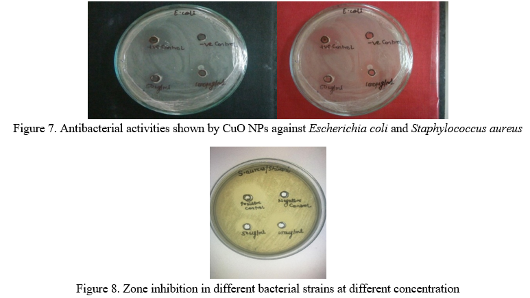
Comparing the antibacterial activities of copper oxide nanoparticles against pathogenic bacteria of gram positive (Staphylococcus aureus) and gram-negative strain (Escherichia coli) as shown in Table 5, it was clearly seen that the synthesized copper oxide nanoparticles showed better antibactericide for Escherichia coli than Staphylococcus aureus..
Conclusion
In this study entitled “Green synthesis, characterization and antibacterial activity of copper oxide nanoparticles using Annona muricata stem extract’’, copper oxide nanoparticles were successfully synthesized using Annona muricata stem extract which provides cost effective, easy and proficient way for synthesis of CuO NPs. The synthesized CuO NPs were characterized by UV-Vis, FTIR, SEM, XRD and its antibacterial activity was investigated. The formation of copper oxide nanoparticles using copper sulphate salt was confirmed by UV-Visible spectroscopy. All the functional groups, bending vibrations and stretching bonds in the sample were confirmed by Fourier Transform Infra-Red (FTIR) spectroscopy. The crystalline nature of the particle was analyzed with the help of XRD (X-ray diffraction) and the size of the particles was calculated using Debye-Scherrer’s equation. The particle size was determined to be in the range of 86-140nm. Scanning Electron Microscopy (SEM) analysis showed that the CuO NPs had almost spherical shape and the average particle size was 100 nm. These results shows that Annona muricata stem extract is capable of producing copper oxide nanoparticles. Agar well method was used to investigate the antibacterial activity of the synthesized nanoparticles. The CuO NPs were found to be highly stable with significant antibacterial activity. The antibacterial activity of the synthesized CuO NPs performed against E. coli and Staphylococcus aureus demonstrated that E. Coli had maximum Zone of inhibition than the gram-positive bacteria, S.aureus.
References
[1] Ceuvas, R., Duran, N., Diez, M.C., Tortella, G.R and Rubilar, O. (2015) Extracellular biosynthesis of copper and copper oxide nanoparticles by Stereum hirsutum, a native white-rot fungus from Chilean forests. Journal of Nanomaterials,4:1-7. [2] Chattopadhyay, D.P. and Patel, B.H. (2012) Preparation, characterization and stabilization of nano-sized copper particles. International Journal of Pure and Applied Sciences and Technology,9:1-8. [3] Gajalakshmi .S., Vijayalakshmi s. and Devi Rajeswari v.(2012) Phytochemical and pharmacological properties of annona muricata: A review. International journal of pharmacy and pharmaceutical sciences.4(2), 3-6. [4] Gregory, V.WII., Petra, K., Ryan, M.B., Jacob, D.M., William, M. A., Delphine, D. and Christopher, L. K. (2012). Green Synthesis of Robust, Biocompatible Silver Nanoparticles Using Garlic Extract. Journal of Nanomaterials. [5] Karthikarani, S. and Suresh, G. (2017) Green synthesis, characterization and its antibacterial activity of copper oxide Nanoparticles using Ocimum sanctum leaves and Piper nigrum extract. International Journal for Research in Applied Science and Engineering Technology,5: 411-419. [6] Koch, R., Zablowski, P., Spreinat, A. and Antranikian, G. (1990). Extremely thermostable amylolytic enzyme from the archaebacterium Pyrococcus furiosus. FEMS Microbiology lettres,71:1-2. [7] Kumar, A., Kaur, K. and Sharma, S. (2013) Synthesis, characterization and antibacterial potential of silver nanoparticles by Morus nigra leaf extract. Indian Journal of Pharmaceuticals and Biological Research,1(4): 16-24. [8] Kumar, P.P.N.V., Shameem, U., Kollu, P., Kalyani, R.L. and Pammi, S.V.N (2015) Green synthesis of copper oxide nanoparticles using Aloe vera leaf extract and its antibacterial activity against fish bacterial pathogens. BioNanoscience ,5 (2): 250-255. [9] Luna, I.Z., Hilary, L.N., Chowdhury, A.M.S., Gafur, M.A., Khan, N. and Khan, R.A. (2015) Preparation and characterization of copper oxide nanoparticles synthesized via chemical precipitation method. Open access library journal,2: e1409 [10] Mude, N., Ingle, A., Gade, A. and Rai, M. (2009) Synthesis of silver nanoparticles using callus extract of Carica papaya-A first report. Journal of Plant Biochemistry and Biotechnology, 18: 83-86. [11] Moghadamtousi S.Z, Goh B.H, Chan C.K., Shabab T., Kadir H.A.(2013) Biological Activities and Phytochemicals of Swietenia macrophylla King. Molecules; 18(9):10465-10483. https://doi.org/10.3390/molecules180910465 [12] Nasrollahzadeh, M., Sajadi, S.M. and Maham, M. (2015) Tamarix gallica leaf extract mediated novel route for green synthesis of CuO nanoparticles and their application for N-arylation of nitrogen containing heterocycles under ligand free conditions. Royal Society of Chemistry Advances,5:40628-40635. [13] Patel, S. and Patel, J.K. (2016) A review on a miracle fruits of Annona muricata. Journal f Pharmacognosy and Phytochemistry.5:137-148. [14] Perumal, K. (2017) Preparation of silver Nanoparticles by Garcinia Mangostana stem extract and investigation of the antimicrobial properties. Biotechnology Research and innovation:23-29. [15] Phiwdang, K., Suphankij, S., Mekprasart, W. and Pecharapa, W. (2013) Synthesis of CuO nanoparticles by precipitation methods using different precursors. Energy Procedia,34:740-745. [16] Prasad, K.S., Patra, A., Shruthi, G. and Chanda, S. (2017) Aqueous extract of Saraca indica leaves in the synthesis of copper oxide nanoparticles: Finding a way towards going green. Journal of Nanotechnology,10: 610-616. [17] Rajendran, A., Siva, E., Dhanraj, C. and Senthikumar S. (2018) A green and facile approach for the synthesis of copper oxide nanoparticles using Hibiscus rosa-sinensis flower extract and its antibacterial activity. Journal of Bioprocessing and Biotechniques,8: 320-324. [18] Rukenya, Z. M., Mbaria, J. M., Mbaabu, M. P., Kiama S. G., Okindo R. O. (2014). Antimicrobial activity of aqueous and methanol extract of naturally growing and cultivated Aloe turkanensis. The Journal of Phytopharmacology,3(5): 343-347. [19] Sadia, S., Arifa, T., Ayyaba, A. and Yongsheng, C. (2016). Plant Mediated Green Synthesis of CuO Nanoparticles: Comparison of Toxicity of Engineered and Plant Mediated CuO Nanoparticles towards Daphnia magna. Nanomaterials,6: 205. [20] Singh, A., Jain, D., Upadhyay, M.K., Khandelwal, N. and Verma, H.N. (2010) Green synthesis of silver nanoparticles using Argemone Mexicana leaf extract and evaluation of their antimicrobial activities. Digest Journal of Nanomaterials and Biostructures,5 (2):483-489. [21] Sivraj, R., Rahman, P.K.S.M., Rajiv, P., Narendhran, S. and Venckatesh, R. (2014) Biosynthesis and characterization of Acalypha indica mediated copper oxide nanoparticles and evaluation of its antimicrobial and anticancer activity. Spectrochimica Acta part A: Molecular and Biomolecular Spectroscopy, 129: 255-258. [22] Thamer, N.A., Muftin, N.Q. and Al-Rubae, S.H.N. (2018). Optimization properties and characterization of green synthesis of copper oxide nanoparticles using aqueous extract of cordia myxa l. leaves. Asian Journal of Chemistry,30:1559-1563. [23] Vasudev D. K. and Pramod S. K. (2013) Green Synthesis of Copper Nanoparticles Using Ocimum Sanctum Leaf Extract . International Journal of Chemical Studies,1(3):1-4. [24] Xia, Y., Xiong, Y., Lim, B. and Skrabalak, S.E. (2008) Shape controlled synthesis of metal nanocrystals: simple chemistry meets complex physiscs. Angewandte Chemie International Edition,48: 60-103. [25] Yoon, K.Y., Byeon, J.H., Park, J.H. and Hwang, J. (2007) Susceptibility constants of Escherichia coli and Bacillus subtilis to silver and copper nanoparticles. Science of Total Environment, 373: 572-575. [26] Yugandhar, P., Vasavi, T., Devi, P. U. M. and Savithramma, N. (2017). Bioinspired green synthesis of copper oxide nanoparticles from Syzygium alternifolium (Wt.) Walp: characterization and evalution of its synergistic antimicrobial and anticancer activity. Applied Nanoscience, 7:417-427. [27] Zhang, li., Yuan, F., Zhang, X. and Yang, L. (2011) Facile synthesis of flower like coper oxide and their application to hydrogen peroxide and nitrile sensing. Chemistry Central Journal, 5:75. [28] Zhang, H., Li, Q., Lu, Y., Sun, D., Lin, X., Deng, X., He, N. and Zheng, S. (2005) Biosorption and bioreduction of diamine silver complex by Corynebacterium. Journal of Chemical Technology and Biotechnology, 80: 285-290.
Copyright
Copyright © 2022 Shivani Kushwaha, Dr. Poonam Prakash. This is an open access article distributed under the Creative Commons Attribution License, which permits unrestricted use, distribution, and reproduction in any medium, provided the original work is properly cited.

Download Paper
Paper Id : IJRASET39498
Publish Date : 2021-12-17
ISSN : 2321-9653
Publisher Name : IJRASET
DOI Link : Click Here
 Submit Paper Online