

Ijraset Journal For Research in Applied Science and Engineering Technology
Improved Accuracy of Brain Tumor Detection Using VGG16
Authors: Asmita Chowdhury, Dharmi Bhimani, Tamanna Choithani, Sejal Thakkar
DOI Link: https://doi.org/10.22214/ijraset.2022.47056
Certificate: View Certificate
Abstract
Brain tumor is a very serious problem of human life. In recent years, brain tumors have become one of the leading causes of death among people. It is difficult to identify the tumor itself. Direct detection and classification of brain tumors has the potential to achieve high efficiency and high levels of prognosis. However, it is well known that the accuracy of automatic identification and classification techniques varies from one technique to another and depends on the nature of the image. For the diagnosis and classification of brain tumors, MRI images have been very useful in recent years. MRI images allow us to diagnose brain tumors. This paper highlights the techniques of CNN and has worked upon VGG16 model.
Introduction
I. INTRODUCTION
Nowadays, the computer cutting edge technology totally relies upon the machine learning techniques such as algorithms and the concepts of statistics which does the role of reinforcing the functional as well as computational aspects (Wei Jin, 2003). In this century, there are myriad of datasets available in the market, which has resulted a sudden rise in the field of Machine learning and Artificial Intelligence. Areas from medical to navy everywhere there is somewhat use of these surging technologies and the demand for which is escalating too. The main advantage of Machine learning is that the system learns and ameliorates just by its experience without being operated by another person (Ayon Dey,2016). Deep Learning, a subset of Machine learning sheds light upon the images by representing them in the form of various pixels thus creating a deep neural network. Its architecture includes RNN, CNN, ANN, deep neural network and much more, which are utilized in numerous fields for image recognition, segmentation as well as computer vision. Deep learning being a comprehensive part of ML usually learns representation of the raw facts. Learning in deep neural networks with conglomerate layers of data does not give the fruitful outcome. In fact, it has already prevailed since neural networks existed, but they were inappropriate at execution. Deep learning enhances the concept that these levels of matter correspond to levels of abstraction or structure. Different column numbers and column sizes can be used to provide different levels of abstraction. Neural networks derive their potrayal using training layers. The primate brain does the equivalent thing as that in the visual cortex, therefore it was anticipated that by utilizing numerous layers in the neural network would permit to learn superior framework (Benuma et al.,2016). A CNN consists of one or more convolutional layers with fully connected layers on top. A compelling class of model, Convolutional neural networks (CNNs) is mostly used for solving the image recognition problems. The proliferation of images and videos on the Internet has led to the development of algorithms in order to analyze the connotation content of images and videos for various uses such as search and summarization (Karpathy et al., 2014). Nowadays, magnetic resonance imaging is very useful in medicine. fields such as medical imaging. Brain tumors are characterized by abnormal tissue growth and uncontrolled cell proliferation, whereby the natural pattern of cell growth and death has failed (Nichal et al.,2017). It is believed that the brain is responsible for controlling emotions, movement, intelligence, speech, memory, emotions, thinking, physical activity, taste, creativity etc. Almost 11,000 people are diagnosed with the brain tumor every year. Therefore, any damage or injury to this vital organ impairs the proper functioning of the human body and will lead to an irregular process. Therefore, it is very important to take care of this precious body. A brain tumor is a rare tumor that involves the uncontrolled growth and reproduction of cells. Brain tumor symptoms depend on the tumor size, type and location. Tumors are malignant and viable threatened by aggressive and limited SP. Depending on the cell type, a brain tumor arises from within or from within the brain. Because of their rapid growth and spread, there are about 130 different types of brain tumors. Tumors vary in appearance, length, shape and presence, so it is arduous to take precise quantifications in order to observe it properly. Computed tomography (CT) scan and Magnetic resonance imaging (MRI) are the advanced imaging techniques accessible to diagnose brain tumors. Therefore, digital image processing plays an important role in medical image analysis for timely and efficient planning. The primary goal of any image processing application for treatment is to use image data such that it isolates the necessary functions that a machine can perform in order to perform a specific test (Mansi Lathera and Dr. Parvinder Singh,2019).
A healthy brain is generally made up of 3 types of tissue: White matter, gray matter and cerebrospinal fluid. The purpose of brain tumor segmentation is detection and identification. Growth of cancerous areas, eg. Active tumor tissue (diffuse or not), tissue necrosis and inflammation (proximal inflammation) ,this is done when abnormal areas are detected compared to normal tissue. Because glioblastomas are infiltrative tumors, their borders are often blurred and difficult to distinguish from healthy tissue. Eyeliner, more than an MRI this method is often used, for example T1 (spin-lattice relaxation). T1 anisotropy (T1C), T2 (spin relaxation), proton density (PD) contrast-enhanced imaging, diffusion-weighted magnetic resonance imaging (DMRI), and fluid subtraction inverse pulse sequence (FLAIR) (Havaei et al., 2016). This paper discusses the technique to detect tumor using Convolutional Neural Network (CNN) model and VGG-16 using the MRI scanned images of the brain.
II. RELATED WORK
Kader et al. published a paper entitled Brain Tumor Detection and Classification on MR Images by a Deep Wave Self-Encoding Model, in which they described the essential implemented steps such as image sharpening, high-pass filter, threshold segmentation, developed seed method and feature extraction are classified based on the deep wavelet auto-encoding model. The proposed model and training tests include BRATS2012, BRAT-S2013, BRATS2014, 2015 Challenge and BRATS 2015 databases. Mean accuracy 99.3%, sensitivity 95.6%, specificity 96.9%, FCPR, 96.9%, FR 5%, FR 5%, 96.9%, FR 5%, 96.9%, FR. 0.031 and JSI 93.3%. Based on the overall test output, segmentation, classification and performance of the proposed DWAE model, it was concluded that the proposed model outperformed 21 existing models published in top-level journals. Their proposed model achieved an excellent overall performance in brain tumor detection and staging, which allows the model to be used in computing strategies for brain tumor detection. The DWAE model demonstrates the importance of deep learning models in the medical field and medical applications.
In a paper titled "Brain Tumor Detection and Classification" by Kanmani and Dr. Pushparani, predicting brain tumors and tumor location the input MRI images are transmitted to their systems as a form of the human brain. After grading tests, they found the correct tumor in the original image. Noise removal and enhancement techniques used in brain MRI scans. The results obtained in this paper were good and effective. The proposed method can be used to detect lung cancer.
In the paper "Brain Tumor Detection and Classification from Multichannel MRI Using Deep Learning and Transfer Learning" by Banerjee et al. present three new CONVNET architectures for invasive brain tumor HGG that perform non-invasively. and LGG, adaptive learning for parallel processing by improving MR tumor imaging and two ConvNet models. A ~12% improvement in classification accuracy was observed on deep ConvNets test data compared to shallow training models. They also found that convolutional networks trained on natural images can perform to their full potential by improving the final transform layers of MRI datasets. In their experiment, we present a method that uses multiple MRI slices containing tumor volume data, which achieves the best accuracy rate of 97.19%. Therefore, it can be concluded that deep convolutional networks can serve as an alternative to surgical biopsy of brain tissue.
Milletari et al. researched on "CNN: MRI Deep for segmentation of deep brain regions in MRI and ultrasound". This was done using CNN which affects territories and conflicts of neurons in brain region. A neural network (CNN) achieved an overall accuracy of 91.3% and a recall of 88%, 81%, and 99% in identifying meningitis, glioma, and pituitary tumors, respectively. A deep learning architecture using a 2D convolutional neural network to discriminate different types of brain tumors from MRI image slices. Techniques such as data assimilation, data preprocessing, premodeling, model optimization, and hyperparameter tuning are used in this paper. In addition, 10-fold cross-validation was performed on the entire data set to check the normality of the model.
The paper published by Fatih Özyurt Eser Sert Engin Avci Esin Dogantekin is titled 'Brain Recognition Based on Convolutional Neural Networks' Neutrosophic Expert Fuzzy Maximum Security Entropy' Elsevier Limited 147. The method used in this paper is based on semi-selection, a strategy that allows for fully automatic selection. Localization and distribution of the anatomy of interest. He also used learning techniques based on hierarchy, dynamic, multi-regional, flexible and adaptable to different situations. different amounts of training data and various measurement data (2D, 2.5D and 3D) are used to predict the final result.
III. DATASET AND METHODOLOGY
A. Dataset
Several methods of detecting tumors in concatenated MRI images were analyzed and applied to the dataset available on the Kaggle website, the images were divided into two groups: with and without tumors, based on which we used various deep learning and machine learning methods and VGG16 model.
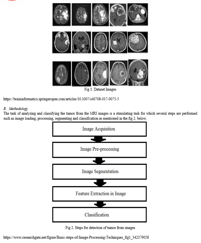
- Data Acquisition: The first step in any image detection is to collect a dataset with numerous images to perform the technique. Next is to load the dataset images. Moreover, the brain tumor dataset is a collection of about 300 images.
- Data Pre-Processing: The crucial preprocessing stage is an image enhancement phase carried out by removal of unnecessary distortion or by displaying clear parts (Kanmani and Dr.Pushparani,2016). Here methods like converting the images into numpy array and performing one-hot coding is carried out. The goal is to reduce noise or redundant detail without introducing excessive distortion, which facilitates downstream analysis. Image processing is the term for processing images at a low level of analysis. This process does not increase the details of the image, but reduces it if the amount of information is irregular. The purpose of this step is to refine the image data by suppressing nonessential artifacts or to enhance image features that are crucial for processing and analysis to a greater extent.
- Image segmentation: The method is the biggest and an essential step for accurate image analysis, apart from this it affects the accuracy of subsequent steps (Patil et al.,2017). Image segmentation is a common technique in image processing and analysis that involves dividing an image into multiple segments or parts, usually based on the characteristics of the pixels in the image.
This is mainly used for:
- Face detection
- Medical imaging
- Machine vision
Image segmentation techniques:
a. Non-Contextual Thresholding
- Simple thresholding
- Adaptive thresholding
- Color thresholding
b. Contextual Segmentation:
- Pixel connectivity
- Region similarity
- Region growing
- Split-and-merge segmentation
c. Texture Segmentation:
- Structural approach
- Statistical approach
- Model based approach
- Filter based approach
4. Feature Extraction in Image: Distinctive features are part of the reduction process, in which the original set of raw data is divided and reduced into smaller parts. The most important characteristic of these large databases is the large number of variables. It helps extract the best features from the data, effectively reducing the amount of data by combining distinct types into features.
5. Classification: The last and final step includes categorizing the image by analyzing it and applying several techniques and concluding whether the MRI image has a tumor or not.
C. Steps
The CNN and VGG16 models are applied on the brain tumor dataset and their performance on classifying the image is analyzed. Steps for the same are:
- Import all the required modules
We imported modules such as tensorflow keras layers , sklearn,numpy,matplotlib etc.
2. Load the image dataset
On loading the images from the dataset ,the output was in the form of two categories mentioned below .
['no', 'yes']
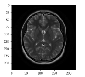
3. Plot an Image
4. Transform the image dataset into numpy arrays
- It converts all the images present in the dataset in a form of array .
5. Applying One-hot encoding to the dataset
- It converts the two categories which were in the text form into numerical form that is yes -1 and no-0.
6. Converting into train and test dataset
- The dataset was splitted into two sets :training set (90%)and test set(10%) .
7. Build the Image Data Generator
- Image data generator simply rotates the image from 0 to 360 degree where the fill mode we have taken as nearest and the rotation range as 15.
8. Build the model
- We have taken base model as VGG16 with weights as imagenet.Then we have applied various layers such as average pooling ,flatten,dense ,dropout.We have calculated activation functions like relu and softmax.
9. Freezing the layers
- It prevents the weights from being modified .
10. Compiling the model
- We have compile the model using adam optimizer with learning rate 0.001,metrics as accuracy and loss as binary cross entropy.
11. Fitting the model
- Gives the summary of the model .
12. Final evaluation of the model
- We have made the predictions using argmax function.
13. Generating the Classification report and Confusion matrix
- We got precision,recall,F1 score,support values for no category as 0.91,1.00,0.95 and 10 respectively. We got precision,recall,F1 score,support values for yes category as 1.00,0.94,0.97 and 16 respectively.
- We received the confusion matrix as [[10 0][1 15]].
14. Accuracy of our model
- The final accuracy of the model is 0.9615.
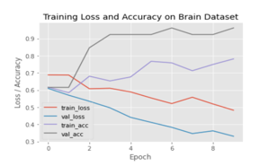
15. Plot the output such as losses and accuracies.
V. FINAL RESULTS
A. Future Scope
Deep learning methods can help classify and categorize brain lesions and reduce the workload of radiologists reading multiple images by prioritizing only the most severe lesions. Over time, this will improve overall efficiency and may reduce diagnostic errors, as deep learning techniques in radiology have achieved equivalent and superior human performance for some pathologies. Spatiotemporal models are commonly used for video classification tasks, which are three-dimensional.(Soumick et al., 2022)
The method structure identifies different tumour regions by highlighting size data for different methods as they show different pathological features while the method structure represents the most mixed part of a tumour and is used to find another part of it.(Ramin et al,2021)
B. Conclusion
In the detection and classification of tumors from brain MR imaging, almost 96% accuracy was achieved .With the above results in mind, we have decided that the approach we propose is different and clearer than normal and unusual, helping clinical professionals to make diagnostic decisions.
Accurate diagnosis of brain tumors remains difficult due to the different appearance, size, shape and structure of the tumor. Although tumor segmentation methods show great potential for the detection and identification of tumors on MRI images, many improvements are still needed to accurately segment and classify the tumor region. Current work presents limitations and challenges in defining the ultrastructure of the tumor region and classifying images.
C. Figures and Tables
Table. Epoch output Table
|
Epoch No. |
Epoch |
|
||
|
Loss |
accuracy |
Val_loss |
Val_accuracy |
|
|
1. |
0.6881 |
0.6164 |
0.6093 |
0.6154 |
|
2. |
0.6869 |
0.5845 |
0.5695 |
0.6154 |
|
3. |
0.6073 |
0.6804 |
0.5344 |
0.8462 |
|
4. |
0.6101 |
0.6530 |
0.4963 |
0.9231 |
|
5. |
0.5888 |
0.6758 |
0.4408 |
0.9231 |
|
6. |
0.5536 |
0.7671 |
0.4115 |
0.9231 |
|
7. |
0.5209 |
0.7580 |
0.3826 |
0.9615 |
|
8. |
0.5578 |
0.7123 |
0.3463 |
0.9231 |
|
9. |
0.5187 |
0.7489 |
0.3621 |
0.9231 |
|
10. |
0.4823 |
0.7812 |
0.3307 |
0.9615 |
References
[1] http://ijcsit.com/docs/Volume%207/vol7issue3/ijcsit2016070332.pdf [2] https://static.googleusercontent.com/media/research.google.com/en//pubs/archive/42455.pdf [3] https://www.sciencedirect.com/science/article/pii/S1877050920306542 [4] https://arxiv.org/abs/1505.03540 [5] https://link.springer.com/chapter/10.1007/978-3-319-75238-9_25 [6] https://pubmed.ncbi.nlm.nih.gov/30733593/ [7] https://pubmed.ncbi.nlm.nih.gov/34573931/ [8] https://www.ncbi.nlm.nih.gov/pmc/articles/PMC7786370/ [9] https://iopscience.iop.org/article/10.1088/1757-899X/1055/1/012115#:~:text=In%20various%20research%20papers%2C%20the,the%20treatment%20to%20the%20patients. [10] https://papers.ssrn.com/sol3/papers.cfm?abstract_id=3867216 [11] https://cis.ieee.org/images/files/Documents/research-grants/Report_SubhashisBanerjee.pdf [12] https://research.nvidia.com/publication/2018-09_3d-mri-brain-tumor-segmentation-using-autoencoder-regularization [13] https://www.hindawi.com/journals/jhe/2022/2693621/alg1/ [14] https://www.researchgate.net/publication/303741861_A_Review_of_Deep_Machine_Learning?enrichId=rgreq-9501680473838212929747b4b8c9728e-XXX&enrichSource=Y292ZXJQYWdlOzMwMzc0MTg2MTtBUzo0NTgzNzkxNzM4NjM0MjRAMTQ4NjI5NzUxOTQxNQ%3D%3D&el=1_x_3&_esc=publicationCoverPdf [15] https://www.researchgate.net/publication/341875705_Research_on_Machine_Learning_and_Its_Algorithms_and_Development [16] https://www.researchgate.net/publication/317547838_A_Review_Paper_on_Brain_Tumor_Segmentation_and_Detection?enrichId=rgreq-9c6fb307b9959a13f65ff1acdc9f5500-XXX&enrichSource=Y292ZXJQYWdlOzMxNzU0NzgzODtBUzo1MTU1NTM5MTM1NjUxODRAMTQ [17] 5OTkyOTAzOTIyNA%3D%3D&el=1_x_2&_esc=publicationCoverPdf [18] https://www.nature.com/articles/s41598-021-90428-8 [19] https://paperswithcode.com/task/brain-tumor-segmentation [20] https://link.springer.com/article/10.1007/s40747-021-00563-y [21] https://www.nature.com/articles/s41598-022-05572-6 [22] https://www.frontiersin.org/articles/10.3389/fnins.2019.00810/full IEEE conference templates contain guidance text for composing and formatting conference papers. Please ensure that all template text is removed from your conference paper prior to submission to the conference. Failure to remove template text from your paper may result in your paper not being published.
Copyright
Copyright © 2022 Asmita Chowdhury, Dharmi Bhimani, Tamanna Choithani, Sejal Thakkar . This is an open access article distributed under the Creative Commons Attribution License, which permits unrestricted use, distribution, and reproduction in any medium, provided the original work is properly cited.

Download Paper
Paper Id : IJRASET47056
Publish Date : 2022-10-11
ISSN : 2321-9653
Publisher Name : IJRASET
DOI Link : Click Here
 Submit Paper Online