

Ijraset Journal For Research in Applied Science and Engineering Technology
Inventive Methodology on Adaptive Hierarchy Segmentation of Bone Cancer Using Neural Network in MATLAB
Authors: Hemant Markhande, Prajwal Selokar, Nisha Raut, Ateef Ahmad, Vijay V Chakole
DOI Link: https://doi.org/10.22214/ijraset.2022.40422
Certificate: View Certificate
Abstract
This research creates a curiosity among people that how technology brings its value to the medical sector. This study is manly based on how Artificial Neural Network (ANN) technology collaborated with the MATLAB and gives magnificent results with the help of Digital X-rays. The project focuses on how technology is creating an ease to the medical sector while diagnosing a patient with a tumor. With this technology doctors can identify and help to cure the tumor at its initial stage. This technology benefited thousands of patients with their tumor. The method is contributing its intelligence to gives more accurate, faster and convenient results. The primary goal of our research is to make an algorithm which brings its value over the table in comparison with the older ones
Introduction
I. INTRODUCTION
Tumor can be defined as a morbid enlargement of body cells which results into abnormal growth of tissue and division of cells itself which turns in swelling of a particular organ. As, medical industry growing day by day. There are different kinds of tumors existing in the human body like brain tumor, bone tumor, lung tumor, etc. While classifying the tumor, image processing is important for analysis. The Outcomes of medical image processing is used to cure the health issues. Bone tumors develop when cells within a bone divide uncontrollably, forming a lump or bloat of abnormal tissue. There are two types of tumors, Benign (Non-cancerous) and Malignant (Cancerous). In our project we are mainly fascinated about image segmentation for a bone image and their classification. In this process input was segmented and the features are extracted in the first module, in the second module the images are classified as Benign or malignant with the use of SVM (Support Vector Machine) and ANN (Artificial Neural Network) classifiers. This project proposed a simple and easy method to detect and classify the bone tumor. X-rays are important medical tool for doctors because x-ray releases the type of radiation called electromagnetic waves, which helps creating pictures of an inner part of the human body in different shades of gray. Digital radiography is widely known as digital x-ray. Digital x-ray images are clearer and help to overcome errors faced by older x-ray technology and that too with quick output, which is helpful for proper diagnosis. This project is to make a digital x-ray based, image processing system which gives a fast and accurate identification of the disease supported by the data gained from the digital x-ray images that are stored in a computer have format like jpg, png etc.
II. METHODOLOGY
A. Morphological Operation
The features of an image processing in the shape or morphology are related with the collection of non-linear operations is called as morphological operation in image processing. Morphological operations create an output image of the same size by applying a structure element to an input image. In a morphological operation, the value of each pixel in the input image with its neighbours is based on a differentiation by corelating with the pixel in the output image. You can construct a morphological operation by selecting the shape and the size of the neighbourhood which is sensitive to certain formation in the input image. The most used morphological operations are dilation and erosion. The morphological operation in an image, erosion clear pixels from object boundaries, whereas the dilation add pixels in the boundaries of an object. The number of pixels added or erased from the objects in an image depending on size, shape of the structured element used to process the image. In the morphological operations erosion as well as dilation, the state of any given pixel in the input image and corresponding pixel are ruled by applying the determined output image. Here, the rule is used to process the pixels define the operation as the erosion or dilation discussed about effective content-based medical image retrieval, an erosion operation is used to shrink or eliminate small objects and command used in MATLAB is the imerode. Dilation operation is used to expand regions and edges and the command used in MATLAB is the imdilate.
B. Dilation
Dilation added pixels in the binary image. There are other types that work on different shades of black and white images. The essential result of the operator on a binary image, is to bit by bit enlarge the boundaries of regions of foreground pixels. Thus, area of the foreground pixels grows in size whereas holes among those regions become smaller. The dilation takes two pieces of data as an input. The initial image which is to be dilated and the second is a set of coordinate points referred to as a kernel.
- Erosion: The erosion process typically uses a structuring part for reducing and probing the shapes composed in an input image. In a binary image, the output pixel is set to 0, if any of the pixels is put to 0. Pixels far side the image border area unit assumed to be put to 1. For grayscale images, uint8 images have the highest value i.e., 255.
- Opening: Opening uses the same structuring method as dilation. Opening method takes two inputs named as gray level erosion and gray level dilation.
- Segmentation: Segmentation phase mainly concentrates on tumor detection. An image segmentation technique such as thresholding and morphological operations is used and the results are compared. Detection of the tumor segmentation technique is applied to the image. In tumor detection clusters of high intensity pixels are isolated from the x-ray image which forms the tumor image. Some of the features like as area, mean, entropy, standard deviation and can be calculated from the tumor image.
- Thresholding and Morphological Operations: Threshold technique in segmentation is easy and effective. It converts the gray scale image into binary image which contains two values 0 and 1. So, accordingly the threshold values like minimum and maximum are decided to detect tumors. It is very effective with the images which have levels of contrast. That’s why we would take the bone tumor images with the high contrast. The threshold value is based on the maximum pixel value in the image, SVM is a tool for machine learning i.e., based large margin data classification. The tool has well-founded classification’s algorithms with good performance. Standard implementation, though provide good accuracy for classification but they are slow. Hence, they can't be applied to a wide range of data mining applications.
- Super-Pixel Segmentation: Super-Pixel segmentation is used to increase the accuracy in the segmentation, to remove the other tissues which are segmented in addition to the tumors. Therefore, in order to get the appropriate outcomes, we have to include additional data for removing other tissues.
C. Proposed Work
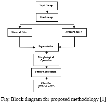
- The algorithm has two stages, initial stage is pre-processing of given x-ray image which segmentation then performs morphological operations.
- Pre-processing is a first step, it designed to boost the quality of images by removing noise, reducing artifacts and increasing contrast to make the segmentation step precise.
- There are several pre-processing techniques such as image adjustment, filtering and histogram equalization etc. Filtering is one among the foremost vital pre-processing techniques because it sharpens the edge of an object and it also help to reduce noise, preserves the edges and smooth unsound images created by the MR imaging system.
- We elect the average filter and bilateral filter. Give x-ray image of bone as input. Convert it to gray scale image.
- Apply average and bilateral filter for noise removal by scrutiny average filter, bilateral contains high noise removal and enhance the quality of image then compute threshold segmentation.
- Compute the morphological operation the output will be a tumor region. For many years, radiologist detects tumor manually, as bone MRI images are complicated and tumors can only be identified by expert physicians with the rapid development of computer technology, computers have become an integral part in medical image acquisition, enhancement, segmentation, labelling and analysis.
- The knowledge and experience of the examiner bears the manual examination of images usually produces highly dependent subjective results.
- Computerized analysis and interpretation of medical images are extraordinarily time and cost effective, and hopefully there are unbiased and reproducible results to be obtained. This let on the comparison of results to be autonomous via human bias and error.
- For in depth understanding of images quantitative information can be beneficial, and which in the form of raw or enhanced image cannot be immediately apparent to the medical examiner. With the scientific approach this result will be obtained more effectively.
D. Block Diagram
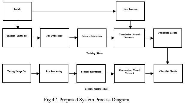
- Neural Network: ANN is propelled by the human brain and it can be utilized for machine learning and counterfeit insights. ANN consists of three layers i.e., (1) the input layer, (2) the hidden layer and (3) the output layer. A neural network method starts once an input data is passed to it. Data is then processed via its layers to produce the specified output. The input or the initial layer of a neural network is made up of artificial neurons the initial data is then fed to the system for processing through the subsequent layers of artificial neural network. The second layer known as hidden layer. It deals with accuracy of results wherever the data must be separated non- linearly. The last layer of an artificial neural network is an output layer. It's accountable for producing the desired result. Here, we trained our algorithm via the neural network for getting the desired output.
- Image Processing: Sometimes the capture image is of poor quality or blurriness in it. Image processing applied in digital pictures. It helps to enhance the quality of an image by de-noising in order to receive improved quality image to take out some useful data. Image processing is like signal processing where input is an image and output are the features associated with that image.
a. Importing the image using optical scanner or digital radiography.
b. Analyzing and manipulating the image which have data compression, image enhancement and spotting patterns that are not seen by human eye like satellite image.
c. Output is the final stage in which result is improved picture or report i.e., based on image analysis.
Conclusion
This proposed system of bone tumor detection with super pixel segmentation is carried out with MATLAB. Also, the detection of brain cancer is carried out with the given set of images. The proposed system is especially dedicated for brain tumor detection. The same system can be further extended to identifying the stages of cancer. In this project we have proposed and plan an exceptional strategy for the location of the bone tumor for various modalities. Our work will be reached out for structuring tumor acknowledged with the mean to decrease the computational time and cost. This strategy gives a sophisticated approach to picture handling operation with examination to find the bone tumor in human body.
References
[1] Integrated approach for bone tumor detection from MRI scan imagery https://ieeexplore.ieee.org/abstract/document/6340479 2017 [2] A Novel Approach for Detecting the Tumor Size and Bone Cancer Stage Using Region Growing Algorithm ttps://ieeexplore.ieee.org/document/7546088/ 2016 [3] Bone Cancer Detection from MRI Scan Imagery Using Mean Pixel Intensity https://ieeexplore.ieee.org/document/7079289/ 2015 [4] A study of UWB imaging for bone cancer detection https://ieeexplore.ieee.org/abstract/document/6340479/ 2012 [5] M.WelsB. .Kelm (2012) “Multi-Stage Osteolytic Spinal Bone Lession Detection from Ct Data with Internal Sensitivity Control”, Proceedings of Medical Science, vol .12, no.5 [6] P. Sinthia, Dr. K. Sujatha, and M. Malathi (2015) “Wavelet Based Decomposition and Approximation for Bone Cancer Image”, Proceedings of Basic and Applied Sciences and Biomedical Engineering, vol. 23, no. 3, pp. 344-350. [7] Shukla S.P. and GulhareKajal Kiran (2013), “Review of Intelligent Techniques Applied for Classification and Pre-processing of Medical Image Data”, IJCSI International Research in Computer Science, vol. 10, no. 3, pp.267-242. [8] Abdulmuhssin Binhssan (2015) “Enchondroma Tumor Detection”, International Proceedings of Advanced Research in Electronics and Communication Engineering, vol. 4, no. 6, pp. 1-4. [9] Markus Harmsen, Benedikt Fischer, Hauke Schramm and Thomas Seidl (2013), “Support Vector Machine Classification Based on Correlation Prototypes Applied to Bone Age Assessment”, Proceedings of Biomedical and Health Informatics, vol. 17, no. 1, pp 190-197 [10] Hubert H. Chuang, Beth A. Chasen and TinsuPan(2013) “Bone Cancer Detection from MRI Scan Imagery Using Mean Pixel Intensity\", Proceedings of Advanced Research in Computer and Software Engineering , vol.1 , no .5, pp .1-7. [11] K. Jalal Deen and Dr. R. Ganesan, “An automated lung cancer detection from CT images based on using artificial neural network and fuzzy clustering methods”, International journal of applied engineering research, 9(22), 17327-17343,2014. [12] Krupali D. Mistry, Bijal J. Talati, “An approach to detect bone tumor using comparative analysis of segmentation technique”, International journal of innovative research in computer and communication engineering, 4(5), 2016. [13] Bhagyashri G. Patil, “Cancer Cells Detection Using Digital Image Processing Methods”, International Journal of Latest Trends in Engineering and Technology, 3(4), 2014. [14] Amit Verma and Gayatri Khanna, “A Survey on Digital Image Processing Techniques for Tumor Detection”, Indian journal of science and technology, 9(14), 2016. [15] Glenn w. Milligan, S. C. Soon, Lisa m. sokol. “The effect of cluster size, dimensionality, and the number of clusters on recovery of true cluster structure”, IEEE transactions on pattern analysis and machine intelligence, 5(1).
Copyright
Copyright © 2022 Hemant Markhande, Prajwal Selokar, Nisha Raut, Ateef Ahmad, Vijay V Chakole. This is an open access article distributed under the Creative Commons Attribution License, which permits unrestricted use, distribution, and reproduction in any medium, provided the original work is properly cited.

Download Paper
Paper Id : IJRASET40422
Publish Date : 2022-02-19
ISSN : 2321-9653
Publisher Name : IJRASET
DOI Link : Click Here
 Submit Paper Online