

Ijraset Journal For Research in Applied Science and Engineering Technology
Melonoma Identification By Using 3D Convolutional Neural Network
Authors: Nkumaran ., Chimmiri Venkata Manoj Kumar, Pothuri Ajay Kumar
DOI Link: https://doi.org/10.22214/ijraset.2022.40816
Certificate: View Certificate
Abstract
Skin biopsy histopathological investigation is one of the essential techniques utilized for pathologists to survey the presence and weakening of melanoma in clinical. A complete and dependable neurotic examination is the consequence of accurately sectioned melanoma and its connection with harmless tissues, and in this way giving precise treatment. In this review, we applied the profound convolution network on the hyper ghastly pathology pictures to play out the division of melanoma. To utilize unearthly properties of three layered hyper otherworldly information, we proposed a 3D completely convolutional network named Hyper-net to section melanoma from hyper phantom pathology pictures. To upgrade the awareness of the model, we made a particular alteration to the misfortune work with wariness of misleading negative in conclusion. The presentation of Hyper-net outperformed the 2D model with the exactness more than 92%. The bogus negative rate diminished by almost 66% utilizing Hyper-net with the adjusted shortfall work. These discoveries showed the capacity of the Hyper-net for helping pathologists in analysis of melanoma in view of hyperspectral pathology pictures.
Introduction
I. INTRODUCTION
Conventional skin tests are recommended by dermatologist to perceive the skin illness in their hidden stages. Therefore, to help this cycle, we proposed a convenient application that can perceive the spot ofthreatening improvement furthermore demand . We proposed a convolutional frontal cortex affiliation and completed two models - Modified Inception model and Modified Google's MobileNet with move learning. The appraisal of the proposed technique is done using HAM10000 dataset which is a variety of multi-source dermatoscopic pictures of ordinary pigmented skin bruises. The exploratory outcomes shows that changed origin model performs better compared to Google's MobileNet.The point is to cultivate a business flexible application to recognize the potential outcomes of earlycompromising improvement with the objective that a real treatment can be proposed to the eccused person.This is on the grounds that an aggregate and solid over the top assessment is the inevitable result of right division of thedefame tissue and its advantage with the innocuous tissue; plus, these examination is the clarification of wary .
Research shows that the most experienced doctors can determine malignant growth to have 79% precision while 91% right conclusion is accomplished ML.In this contextual analysis our errand is detecyt 3 mos normal kinds of diseases at beginning phase utilizing cnn
There are 3 primary kinds of malignant growths
- Basil skin malignant growth
- Squamous skin cell malignant growth
- melonama malignant growth
?n this rousing and empowering in pathology picture examination, which covers a wide scope of utilizations including clinical picture characterization, location and division for a variety of diseases.In terms of division, the errand expects grouping to be performed on every pixel of the picture. Long et al. proposed an original methodology which supplanted completely associated layers by completely convolutional layers with the goal that the organization can yield a total division picture in a solitary forward.
Nonetheless, as the convolutional channels extricate more unique highlights layer by layer, the goal of info picture diminishes slowly. To protect the underlying goal, the most common engineering named U-net was proposed, which assembles an encoding way and a symmetric unraveling method for showing up at careful objective.
II. LITERATURE SURVEY
Despite recent improvements in prevention, diagnosis, and treatment, vast differences in melanoma burden still exist between populations. To assess global, regional, and national melanoma incidence, mortality, and disability-adjusted life year estimates from the Global Burden of Disease in the year. Total prevalence was divided into four disease phases and multi-plied with disability weights to generate years lived with disability In Africa; mortality rates were greater in males than females. The highest death rate is in the age of 70. GBD results can help shape melanoma research and public policy. This article is protected by copyright.
Skin cancer, the most common human malignancy, is primarily diagnosed visually; beginning with an. automated classification of skin lesions using images is a challenging task owing to the fine-grained variability in the appearance of skin lesions. Deep convolutional neural networks (CNNs) show potential for general and highly variable tasks across many fine-grained object categories. Here we demonstrate classification of skin lesions using a single CNN, trained end-to-end from images directly, using only pixels and disease labels as inputs. We test its performance against 21 board-certified dermatologists on biopsy-proven clinical images with two critical bi-nary classification use cases: keratinocyte carcinomas versus benign. The first case represents the identification of the most common cancers; the second represents the identification of the deadliest skin cancer...
III. PROPOSED SYSTEM
We applied the significant convolution network on the hyper spectral pathology pictures to play out the division of melanoma. To use spooky properties of three layered hyper spectral information, we proposed a 3D totally convolution network named Hyper-net to section melanoma from hyper spectral pathology. To work on the consciousness of the model, we made a specific acclimation to the mishap work with care of misdirecting negative in assurance. We applied the significant convolution network on the hyper spectral pathology pictures to play out the division of melanoma. To use spooky properties of three layered hyper spectral information, we proposed a 3D totally convolution network named Hyper-net to piece melanoma from hyper spectral pathology. To work on the attention to the model, we made a specific acclimation to the hardship work with care of deceiving negative in assurance.
A. Benefits
- False negative rate lessened
- The execution of Hyper-net outflanked the 2D model with the precision over 92%. we can investigate re-partner testing task incorporating greater dataset with different degrees.
B. Ex?st?ng System
Our past work has utilized the article based multistage discovery technique to investigate on this subject with a little dataset. With the quickly developing procedure, we can explore seriously testing task including bigger dataset with various extents. In this we focus on the on-going picture managing strategy executed in skin biopsy masochist evaluation, to the degree that its real breaking point in giving objective norms and further making proficiency for clinical screening.
C. Disadvantages
- High-accuracy imaging procedures and computational power, have as of now given advancement in the field of clinical conclusion It examination just modest quantity of dataset.
- The misfortune work with wariness of bogus negative in conclusion is more
IV. IMPLEMENTATION
A. Systemarch?cture
UPLOAD DATA
LOAD DATASET
PREPROCESSOR DATASET
DATA ANALYSIS
MODEL TRAINING AND EVALUATION
MODEL INTERPRETATION
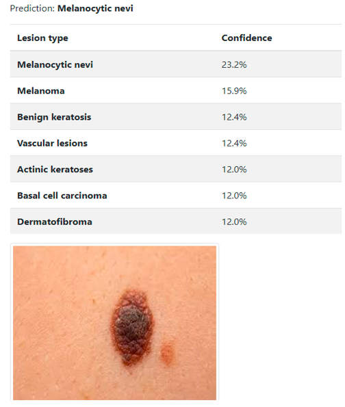
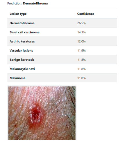
PREDICT MELONOMA
B. Programming Requirements
The practical prerequisites or the general description records incorporate the item perspective what's more, features, working system and working environment, representations necessities, plan constraints and client documentation.
C. Equipment Neccesities
- FESABILITY Study: The reasonableness of the endeavour is analyzed in this stage and key understanding is progressed with a very wide game plan for the endeavour and a couple of statements. During system examination the believability examination of the proposed structure is to be finished...This is to ensure that the proposed system isn't a load to the association
a. Economical attainability
b. Technical feasibility
c. Social feasibility
2. Financial FESABILITY: This study is completed to check the monetary effect that the framework will have on the organization. How much asset that the organization can fill the innovative work of the framework is restricted? Along these lines the created framework too affordable and this was accomplished in light of the fact that a large portion of the advances utilized are unreservedly accessible. Only the altered things should be purchased.
3. Specialized FESABILITY: This study is done to check the specialized attainability, that is to say, the specialized prerequisites of the framework. Any framework created should not have an appeal on the accessible specialized assets. This will prompt high requests being put on the client. The created framework should have an unassuming requirement, as just insignificant or invalid changes are required for carrying out this framework.
4. Social FESABILITY: The part of study is to check the degree of ac-acceptance of the framework by the client. This incorporates the most common way of preparing the client to utilize the framework proficiently. The client should not feel compromised by the framework, rather should acknowledge it as a need. The degree of acknowledgment by the clients exclusively depends on the strategies that are utilized to educate the client about the framework and to make him acquainted with it. His degree of certainty should be raised so he is additionally ready to make a few con-structure analyses, which is invited, as he is the last client of the framework.
D. Module Description
- Upload dataset Using this module dataset is uploaded here.
- Read data Using this module data set is read.
- Loading the data Using this module data is loaded.
- Data analysis Using this module data analysis is take place.
- Model training and evaluation Using this module model training and evaluation takes place.
- Model interpretation Using this module model interpretation is build.
- Predict melanoma Using this module skin diseases is predicted here.
E. Algor?thm
- CNN: We applied the profound convolution network on the hyperspectral pathology pictures to play out the division of melanoma. To utilize ghastly properties of three layered hyperspectral information, we proposed a 3D completely convolutional network named Hyper-net to fragment melanoma from hyperspectral CNN: In profound learning a convolutinal brain network is propelled by natural cycle in which the availability design among neurons and neurons looks like the association of the creature visual cortex. It is an AI work that mirrors the working of human mind in handling information for use in distinguishing the articles perceiving the speech,translating dialects and making decisions.To recommend this strategy writer previously depicting idea to carry out different models in which one model can identify or perceive human transcribed digits and second model can recognize opinion from message sentences which can be given by human about government. In our expansion model we added one more model which can identify feeling from individual face picture. Individual face appearances can depict opinions better than words or sentences. So our augmentation work can foresee feelings from individual face pictures. To show how to construct a convolutional brain network based picture classifier, we will assemble a 6 layer brain network that will distinguish and isolate one picture. This network that we will fabricate is a tiny organization that we can run on a CPU also However, our goal is to tell the best way to construct a genuine world convolutional brain network utilizing TENSORFLOW. On the off chance that you stack neurons in a solitary line, it's known as a layer; which is the following structure square of brain organizations. See beneath picture with layers
- Input Layer: It should contain the image data.Its generally represents the input layer of he image.
- H?dden Layer: The hidden layers of a CNN typically consist of convolutional layers, pooling layers, fully connected layers, and normalization layers. Here it simply means that instead of using the normal activation functions defined above, convolution and pooling functions are used as activation functions.
- Output Layer: The output layer in a CNN as mentioned previously is a fully connected layer, where the input from the other layers is flattened and sent so as the transform the output into the number of classes as desired by the network
F. Hyperspectral Patholoy Image Preprocess?ng
There some compels in information obtaining and execution, for example, the outflow spectra of the enlightenment sources, the transmission of the optics in the magnifying lens and the identification responsiveness of the charge coupled gadget (CCD) camera, bringing about excess and uproarious information somewhat. We followed the band choice system in [50] involving shared data as the pointer. The appraisal of common data between each band and the reference band. M I n(Hn, R) = H P(Hn, R) ∗ log P(Hn, R) P (Hn) ∗ P(R) (1) where n ∈ [1, 60] addresses the nth band, P() addresses the likelihood appropriations of dark size of the nth band. Additionally, an alignment cycle is required in advance, expected to dispense with boisterous information and acquire the huge trademark spectra of histopathology . particularly for biomedical applications, the normal strategy is to utilize the at the foreordained frequencies by imaging a coverslipped slide containing no segment. . The adjustment is determined by condition , where R, B and D addresses the crude picture, the clear picture and the dim picture, separately.
V. RESULT ANALYSIS
Conclusion
Our work demonstrated that the 3D convolu-tional neural network can be used to segment melanoma in hyperspectral pa-thology images, therefore assisting pathologists in determining melanoma deterioration. Melanoma can be identified from healthy tissue under both 10X and 20 X magnifications with accuracy more than 92%. Also, to improve the sensitivity of di-agnosis, we proposed the Hyper-net neural network by making specific adjustment of misfortune work, prompting upgraded execution of incredibly re-diction in bogus positive and misleading negative pre-phrasings., we showed that hyper spectral pathology pictures contained rich instructive properties of tissues to work on the segmentation results and guarantee analy-sis. Although the accuracy was reasonable, our work only covered limited diversity of tissues in skin pathology. In addition, there are many other complex and rare tissues that may need pathologists’ pro-fissional knowledge such as cyst, necrosis and inflammation. In terms of our MHSI system, we intend to upgrade the current MHSI system to enlarge the spectra range so as to include spectra between 400 and 750, and to consider more significant features to enhance the quality of channel. Last but not least, we have to admit that there is a long way before the hyper spectral dad thology framework could be utilized in clinical that are the reason scientists couldn\'t quit investigating different methods of likelihood. Clearly, profound organizations opened another understanding into clinical field with watchfulness and thought on clinical situations.
References
[1] Karimkhani , “The burden global of melanoma: Results from the global burden of disease study 2015,” Brit. J. Dermatol., vol. 177, no. 1, pp. 134–140, Jul. 2017. [2] A. Esteva et al., “Dermatologist-level classification of skin cancer with deep neural networks,” Nature, vol. 542, no. 7639, p. 115, 2017. [3] A. M. Glazer, R. R. Winkelmann, A. S. Farberg, and Ds.Rigel, “finds the incidence of melonama in us . JAMADermatol vol. 153, no. 2, pp. 225–226, 2017. [4] S. W. Menzies et al., “Dermoscopic evaluation of amelanotic and hypomelanotic melanoma,” Arch. Dermatol., vol. 144, no. 9, pp. 1120–1127, 2008.
Copyright
Copyright © 2022 Nkumaran ., Ch?mm?r? Venkata Manoj Kumar, Pothur? Ajay Kumar . This is an open access article distributed under the Creative Commons Attribution License, which permits unrestricted use, distribution, and reproduction in any medium, provided the original work is properly cited.

Download Paper
Paper Id : IJRASET40816
Publish Date : 2022-03-16
ISSN : 2321-9653
Publisher Name : IJRASET
DOI Link : Click Here
 Submit Paper Online