

Ijraset Journal For Research in Applied Science and Engineering Technology
Prediction of Pneumonia Using CNN
Authors: Anuradha Kodali, Vanamala Daniel, Dasari Raviteja, Sai Charan Atelly, Aditya Sagar, Shagvi
DOI Link: https://doi.org/10.22214/ijraset.2022.42110
Certificate: View Certificate
Abstract
Pneumonia is one of the most deadly and dangerous lung infections. Pneumonia is a contagious, deadly disease in the lungs that is caused by bacteria, fungi, or viruses that invade the airways of a person\\\\\\\'s lungs with a load of fluid or pus. Chest X-ray is a common method used to diagnose pneumonia and requires a medical professional to evaluate the outcome of the X-ray. For non-specialists, it is difficult to tell if a patient has pneumonia with chest X-ray images. When a convolutional neural network is used to handle this task, it will improve the diagnosis of pneumonia and reduce the workload of physicians. Medical Imaging has a great scope of application for the latest advances in computation. With emerging computer technology, the development of an automated pneumonia detection system is using Convolutional Neural Networks in line with different standardization (Dropout, L2, and Dropout + L2) and development methods (SGDM, RMSPROP and ADAM). Treatment for this disease is now especially possible, if the patient is in a remote area and medical services are limited. Therefore, the idea is to create an algorithm that automatically detects whether a patient has pneumonia or not by looking at chest X-ray images.
Introduction
I. INTRODUCTION
Medical Imaging has a great scope of application for the latest advances in computation. Convolutional Neural Networks (CNN's) has been a major success for image-based analysis. They can be used effectively to separate images. Over the past decade, there have been remarkable achievements in the medical field, especially in the medical field. Advanced Reading and CNN are actively used in a variety of medical studies especially those with photographic data. They are used for wound separation, isolation and tumor separation, image enhancement, abnormal diagnosis and disease, nucleus detection, etc. With the advent of transfer learning, new skills have been added to CNN. Pass on learning models like VGG16 etc. trained using image data.
Chest tests that are widely used to detect lumps in the lungs can also be used to diagnose other diseases such as pneumonia, effusion, cardiomegaly etc. Among these, pneumonia is a contagious, deadly disease that affects millions of people, especially the elderly. over 65 and you suffer from chronic diseases such as asthma or diabetes. In the process of diagnosing pneumonia, chest x Radiation is considered the most effective way to determine the severity and location of a septic region in the lungs. However, examining chest radiographs is not a hobby for radiotherapists. In X-ray images of the chest, the appearance of pneumonia may be blurred and poorly understood by some diagnoses. Chest X-Ray tests especially in the case of Pneumonia can be misleading because many other problems such as heart failure, lung scars etc. can imitate pneumonia. This is the main reason for the incorrect classification of X-ray images in the database. Therefore, the task is challenging and the development of an algorithm for diagnosing thoracic diseases such as Pneumonia will increase access to clinical settings in remote areas.
Pneumonia is among the leading causes of death from infection. It is called an infection caused by air sacs in the lungs. There are various causes for it, including various viruses, bacteria, and fungi. It can have moderate to severe effects on people of any age group. Some types of pneumonia can be treated with vaccines. Chest x-ray is a simple, easy and effective method that allows doctors to diagnose 2 internal organs or diagnose abnormalities and diseases of the blood vessels, lungs, heart, airways and bones. Pneumonia, caused by a lung infection, can be diagnosed with a chest xray. In this study, automated models were developed to detect pneumonia on x-rays without human assistance.
A. Existing System
Chest x-rays are hard to interpret without an expert radiologist. A lot of researchers are striving to make x-ray analysis automated.
In the existing system, The Experts were needed to recognize/confirm by seeing the chest x- rays whether the person is suffering with pneumonia disease or not.
These includes expert availability, cost, Time etc.
B. Drawbacks
- Using a Support vector machine algorithm only two diseases can be detected and it only displays whether it is diseased or not.
- K Means for detecting the rice leaf plant diseases will not classify the type of disease
C. Proposed System
A complete solution for the diagnosis of pneumonia on chest X-rays is suggested.
Diagnosis involves a chest x-ray interpreted by a radiologist.
In these types of neural networks are designed to detect pneumonia in x-ray images of the chest. The basic convolutional neural network (CNN) models, VGG16, augmentation are developed using CNN methods.
The models were then trained in a pediatric pneumonia database that had X-Ray images divided into three categories namely General, bacterial pneumonia, Viral pneumonia. We use different network models to find.
So that different models offer different levels of accuracy. Therefore, with these we can also find the best model for adoption.
II. LITERATURE ZREVIEW
Over the past decade, a few machine learning based on automated methods of identifying different types of pneumonia has been extensively researched. [15] Fiszman used a Natural language analysis (NLP) tool for diagnostics related to harmful bacterial pneumonia X-ray disease of the chest. The functionality of this type of comprehensive app application is very efficient highly comparable to a human expert. Chapman et al demonstrated three computer systems use legal basis, possible Bayesian network, and decision drug diagnostic chest X-ray report associated with harmful bacterial pneumonia. A The feasibility study of an NLP-based monitoring program was conducted to identify pneumonia-related health care in newborns. However, effective clinical applications of these types of methods are limited due to the reliance on information extracted from narrative patient reports. Parveen reports an unidentified un-c-path A phased learning algorithm to detect X-ray images of pneumonia. This the method improves the accuracy of the sections as c-incomprehensible means distributing the weights throughout input pixels for X-ray images. Rajpurkar showcased ChexNet, 121 layer depth convolutional neural network (CNN), which provides access to or to identify pneumonia using a temperature map to locate the area of ??infection. Germany introduced a DL-based transfer learning framework for the detection of childhood pneumonia using X-ray images of the chest. However, there are no methods used to differentiate X-ray images from them CS framework pneumonia to meet the need for remote end analysis. Researchers they use the results of machine learning predictions to solve health problems science. A large amount of information can be extracted and used for future prevention dangerous diseases. To extract information from Python language medical pictures is used. High-resolution data contains medical size images number of feature adjectives. To extract multiple feature adjectives, feature extraction techniques used in high-resolution X-ray images. In-depth neural learning networks trained with extruded data to create a guessing model. Research is like that moreover pneumonia was performed using machine learning phases.
Many researchers have encountered the problem of overlapping images accuracy. Rubin developed a CNN model to detect common pre-existing thorax disease 9 and X-ray images of the back chest. The MIIC-CXR database was used for maximum performance automatic detection of these images. The database was divided into training, testing and validation is set as 70%, 20% and 10%, respectively. Additional data and pixels practice has been used to improve overall performance. Their DualNet CNN model obtained a median AUC of 0.72 and 0.688 of PA and AP, respectively.
A deep convolutional neural network to differentiate pulmonary tuberculosis was developed by Lakhani, Transfer learning models like AlexNet and GoogleNet and is used to classify chest X-ray images. The database was divided into training, testing and validation sets as 68%, 14.9% and 17.1%, respectively. Adding data and pre-processing techniques used to obtain the most efficient model AUC of 0.99. The accuracy and memory of the model was 100 and 97.3%. AG-CNN model was developed by Guan to diagnose thorax disease. ChestX-ray14 was used to do so detect thorax in chest X-ray images. Attention to international and local branch CNN was used for partition purposes. Their model was better than other models mentioned in their research paper, they obtained AUC of 0.868.
The deep convolutional neural network model was developed by Rajpurkar to divide chest X-ray images into pneumonia and 14 other diseases. ChestX-ray14 data was used to train the model. They compared their ChXNet (121 layered model) model) and study radiologists. Their ChXNet model earned F1 points (95% CI) of the 0.435 best performing radiologists who scored F1 (95% CI) of 0.387.
A convolutional network model with five-layer conversion followed by layers of multi-layered, three-layer layers that are fully connected and trained by Krizhevsky. This network contained 60 million different parameters. By rent To stop, this model has experienced a 5% 17% error. Simonyan developed a the most accurate model uses multiple kernel-sized filters to achieve the top five 92.7% accuracy test.
This model was trained in the ImageNet database and sent to ILSVRC 2014 competition Convolution Neural Network(CNN) of isolation and separation of the brain tumor MRIs were developed by Xu. Many strategies such as adding data, 10 Select the features and integration techniques used in this model. Verification 474 V. Sirish Kaushik the accuracy of the separation achieved by this model is 97.5%, too segment verification accuracy is 84%, 256 × 256 pixels for front chest size radiographs were provided in a deep convolutional neural network for diagnosis unusual.
A convolutional neural network with five active transition layers ReLU leak, intermediate integration and three fully integrated layers were developed by Anthimopoulos for patterns of interstitial lung disease in a database containing 14,696 photos of seven different classes. This model gained segmentation 85.5% accuracy. Creates a neural residual network (RNN) to separate images available in ImageNet database. RNN introduced the concept of connecting shortcuts to deal with the problem of gradient collapse. This model has been submitted to the ILSVRC 2015 has achieved the accuracy of the modern phase. Transfer learning model, AlexNet extension, using data augmentation techniques, was developed by Glozman. This model was trained on the ADNI website. Two models of neural network have presented by Hemanth namely MCPN and MKNN. These models differentiate MRIs high accuracy and high integration time for Artificial Neural Networks. Neural convolution network of isolation and separation of the brain tumor MRIs were developed by Xu. Many techniques like adding data, feature Selection and integration methods were used in this model. 474 V confirmation. Sirish Kaushik's accuracy in the classification obtained by this model is 97.5%, too segment verification accuracy is 84%, 256 × 256 pixels for front chest size radiographs were provided in a deep convolutional neural network for diagnosis unusual.
A convolutional neural network with five active transition layers ReLU leak, intermediate integration and three fully integrated layers were developed by Anthimopoulos for patterns of interstitial lung disease in a database containing 14,696 photos of seven different classes.
This model gained segmentation 85.5% accuracy. Creates a neural residual network (RNN) to separate images available in ImageNet database. RNN introduced the concept of connecting shortcuts to deal with the problem of gradient collapse. This model has been submitted to the ILSVRC 2015 has achieved the accuracy of the modern phase. Transfer learning model, AlexNet extension, using data augmentation techniques, was developed by Glozman.11
This model was trained on the ADNI website. Two models of neural network have presented by Hemanth namely MCPN and MKNN. These models differentiate MRIs high accuracy and high integration time for Artificial Neural Networks In more recent times, exploring machine learning algorithms (ML) for discovery thoracic diseases have received attention in the field of medical imaging classification.
Lakhani and Sundaram (2017) [17] proposed a way to get lungs tuberculosis following the formation of two different DCNNs AlexNet and GoogleNet. Separation of lung nodules especially in the diagnosis of lung cancer proposed by Huang et al. [11] and used in-depth reading strategies.
The effectiveness of different types of Convolutional Neural Networks (CNNs) for the discovery of abnormalities on X-Rays of the chest were proposed by Islam et al. [16] using publicly available OpenI databases [1]. For the better to test machine learning in chest tests, Wang et al. (2017) [24] published a Large database of X-Rays front chest.
Recently, Pranav Rajpurkar, Jeremy Irvin, et al. (2017) [18] reviewed this database for finding pneumonia at a better level than radioologists, they called their model ChexNet uses DenseNet-121 layer architecture to detect all 14 infections from the many 112,200 images found in the database.
Behind the CheXNet model [18], Benjamin Antin et al. (2017) [2] worked on the same database and proposed editing retreat model for pneumonia. Pulkit Kumar, Monika Grewal (2017) [19] using cascading convolutional networks to present their research multilabel classification of thoracic diseases. Zhe Li (2018) [25] recently proposed a a convolutional network model for diagnostic and local practice.
The proposed system is used to detect pneumonia using a variety of enhancements methods such as SGD, RMSprop, Adam. Several modification models such as VGG16, Augmentation were used to build the model..
III. METHODOLOGY
A. Dataset Collection
Dataset of chest x-ray images is collected. It is divided into different train and test ratios: 90-10, 80-20, 70-30, 60-40 respectively. Train and test consist of three categories namely bacterial pneumonia, viral pneumonia and normal chest x-ray images.
B. Data Preprocessing
Dataset is preprocessed to prevent over fitting, with the parameters like rescale, shear range, zoom range, horizontal flip, flow from directory.
C. Model Building and Evaluation
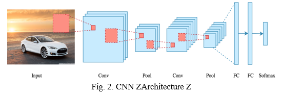
- CNN Algorithm: Today Convolutional Neural Networks(CNN) is one of the go-to solutions for every image-related and image recognition problem. In terms of accuracy, they give the best. It is also successfully applied to recommender systems, natural language processing, medical diagnosis, and more. The advantage of CNN compared to other machine learning algorithms is that it automatically detects the most important features of the image without any human supervision. CNN is also computationally efficient. It uses very special convolution operations and pooling operations and performs parameter sharing by applying different layers. That enables CNN models to be compatible and to run on any device, making them universally attractive. We are dealing with a great and efficient model which performs automatic feature extraction from images to achieve superhuman accuracy (CNN models now do image classification better than humans)
2. CNN Architecture
From the University of Oxford K. Simonyan and A. isserman proposed the VGG16 convolutional neural network (CNN) model in the paper “Very Deep Convolutional Networks for Large-Scale Image Recognition”. In the ImageNet dataset, which contains 14 million photos divided into Z1000 classes, the model achieves 92.7 percent top-5 test accuracy. Through their article, VGG-16 was one of the best models submitted to the 2014 ILSVRC. It outperforms Alex Net by substituting large kernel-sized filters (11 in the first convolutional layer and 5 in the second convolutional layer, respectively) with a large number of 33 kernel-sized filters in a sequential manner.
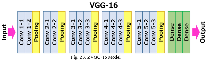
VGG-16 model is mainly composed of three types of layers: convolutional layers, pooling layers, and dense layers. When these layers are stacked, a VGG-16 architecture is created.
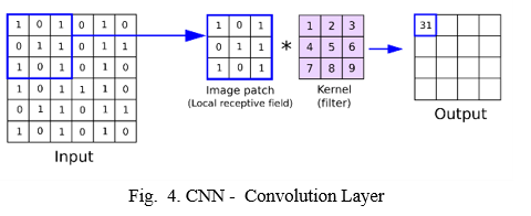
a. Convolution Layer: A convolutional layer is a primary layer that retrieves the different features from the input photos. This layer performs the mathematical process between the input image and a filter of a given size MxM. Sliding the filter across the input image produces the dot product between the filter and the parts of the input image. The output is the Feature map, which contains information about the image's corners and edges.
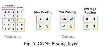
b. Pooling Layer: When we pool an image, we're computing a total value based on all of the values there, so we take a tiny amount of the input and try to average it out, referred to as "average pooling," or pick the maximum value, referred to as "max pooling."
c. Dense Layer: The dense layer in convolutional neural networks is a simple layer of neurons in which each neuron receives input from all of the neurons in the previous layer of the model. In CNN, Dense layers are used to identify images based on convolutional layer output.
D. Implementation and Testing
- Step-1: Open Anaconda Prompt: The first step is to download and install the Anaconda software package.
- Step-2: Anaconda Activate: Activate the Anaconda when you've installed all of the files.
- Step-3: Go to the Project Directory: All project-related files must be kept in one place. Using the Linux commands at the anaconda prompt, navigate to the project directory.
- Step-4: Enter the Command: Because the code is written in the Python programming language, once you've been redirected to the project directory, run the project using the command python filename.py.
E. Web page Results
Our model, which employs CNNs, is deployed on a web page where the user can directly upload the Chest X-rays image to check whether it is diseased or not. If it is affected, it also displays the disease name. The model has an average accuracy of 95.27 percent.

Conclusion
Presence of expert radiologists is the top most necessity to properly diagnose any kind of thoracic disease. The model primarily aims to improve the medical adeptness in areas where the availability of radiotherapists is still limited. It facilitate the early diagnosis of Pneumonia to prevent adverse consequences (including death) in such remote areas. In this classifier, we observed the performance of various pre trained CNN models.
References
[1] 2016. Openi Dataset, 2016, [online] Available: https://openi.nlm.nih.gov. Automatic detection of acute bacterial pneumonia from chest X-ray reports [2] Benjamin Antin, Joshua Kravitz and Emil Martayan, Detecting Pneumonia in Chest X-Rays with Supervised Learning, 2017. [3] CDC Features. Centers for Disease Control and Prevention, Centers for Disease Control and Prevention, 22 Oct. 2018, www.cdc.gov/pneumonia/index.html. [4] D.-F. Wang, Y. Guo, X. Wu, J. Na and G. Litak, \\\\\\\"Planetary-gearbox fault classification by convolutional neural network and recurrence plot\\\\\\\", Appl. Sci., vol. 10, no. 3, pp. 932, Jan. 2020. [5] https://en.wikipedia.org/wiki/Shear_mapping [6] https://stackoverflow.com/questions/57301330/what-exactly-the-shear-do-in imagedatagenerator-of-keras [7] https://www.i2tutorials.com/explain-what-flask-is-and-its-benefits/ [8] https://www.researchgate.net/publication/340961287_Pneumonia_Detection_Using_C onvolutional_Neural_Networks_CNNs#pfd [9] https://www.studytonight.com/post/random-zoom-image-augmentation-keras imagedatagenerator [10] J Am Med Inform Assoc, 7 (6) (2000), pp. 593-604 [11] Kai-Lung Hua, Che-Hao Hsu, Shintami Chusnul Hidayati, Wen-Huang Cheng and Yu-Jen Chen, \\\\\\\"Computer-aided classification of lung nodules on computed tomography images via deep learning technique\\\\\\\", Onco Targets and therapy, vol. 8, 2015. [12] Kermany, D. K., and M. Goldbaum. \\\\\\\"Labeled optical coherence tomography (OCT) and Chest X-Ray images for classification.\\\\\\\" Mendeley Data 2 (2018). [13] Krizhevsky, Alex, Ilya Sutskever, and Geoffrey E. Hinton. \\\\\\\"Imagenet classification with deep convolutional neural networks.\\\\\\\" Advances in neural information processing systems. 2012. [14] Liang, Gaobo, and Lixin Zheng. \\\\\\\"A transfer learning method with deep residual network for pediatric pneumonia diagnosis.\\\\\\\" Computer Methods and Programs in Biomedicine (2019). [15] M. Fiszman, W.W. Chapman, D. Aronsky, R.S. Evans, P.J. Haug 37 [16] Mohammad Tariqul Islam, Md Abdul Aowal, Ahmed Tahseen Minhaz and Khalid Ashraf, \\\\\\\"Abnormality detection and localization in chest x-rays using deep convolutional neural networks\\\\\\\", 2017. [17] Paras Lakhani and Baskaran Sundaram, \\\\\\\"Deep learning at chest radiography: automated classification of pulmonary tuberculosis by using convolutional neural networks\\\\\\\", Radiology, vol. 284, no. 2, 2017. [18] Pranav Rajpurkar, Jeremy Irvin, Kaylie Zhu, Brandon Yang, Hershel Mehta, Tony Duan, Daisy Ding, Aarti Bagul, Curtis Langlotz, Katie Shpanskaya et al., \\\\\\\"Chexnet: Radiologist-level pneumonia detection on chest x-rays with deep learning\\\\\\\", 2017. [19] Pulkit Kumar, Monika Grewal and Muktabh Mayank Srivastava, \\\\\\\"Boosted cascaded convnets for multilabel classification of thoracic diseases in chest radiographs\\\\\\\", International Conference Image Analysis and Recognition, pp. 546552, 2018. [20] Rajpurkar, Pranav, et al. \\\\\\\"Chexnet: Radiologist-level pneumonia detection on chest x-rays with deep learning.\\\\\\\" arXiv preprint arXiv:1711.05225 (2017). [21] Saul, Can Jozef, Deniz Yagmur Urey, and Can DorukTaktakoglu. \\\\\\\"Early Diagnosis of Pneumonia with Deep Learning.\\\\\\\" arXiv preprint arXiv:1904.00937 (2019). [22] Stephen, Okeke, et al. \\\\\\\"An Efficient Deep Learning Approach to Pneumonia Classification in Healthcare.\\\\\\\" Journal of healthcare engineering 2019 (2019). [23] X. Guo, L. Chen and C. Shen, \\\\\\\"Hierarchical adaptive deep convolution neural network and its application to bearing fault diagnosis\\\\\\\", Measurement, vol. 93, pp. 490- 502, Nov. 2016. [24] Xiaosong Wang, Yifan Peng, Le Lu, Zhiyong Lu, Mohammadhadi Bagheri and Ronald M Summers, \\\\\\\"Chestx-ray8: Hospital-scale chest x-ray database and benchmarks on weakly-supervised classification and localization of common thorax diseases\\\\\\\", Computer Vision and Pattern Recognition (CVPR) 2017 IEEE Conference on, pp. 34623471, 2017. [25] Zhe Li, Chong Wang, Mei Han, Yuan Xue, Wei, Li-Jia Li, et al., \\\\\\\"Thoracic disease identification and localization with limited supervision\\\\\\\", 2017
Copyright
Copyright © 2022 Anuradha Kodali, Vanamala Daniel, Dasari Raviteja, Sai Charan Atelly, Aditya Sagar, Shagvi . This is an open access article distributed under the Creative Commons Attribution License, which permits unrestricted use, distribution, and reproduction in any medium, provided the original work is properly cited.

Download Paper
Paper Id : IJRASET42110
Publish Date : 2022-05-01
ISSN : 2321-9653
Publisher Name : IJRASET
DOI Link : Click Here
 Submit Paper Online