

Ijraset Journal For Research in Applied Science and Engineering Technology
Recognition and Classification of Diabetic Retinopathy Using Retinal Fundus Images
Authors: Mr. Anand M, Sanjana B Raj, Chinmayi K S, Harshitha S
DOI Link: https://doi.org/10.22214/ijraset.2022.44594
Certificate: View Certificate
Abstract
Diabetic Retinopathy which impacts the retinal part in the eye because of high sugar degree in the blood. Which reasons retinal damage in the eye leads to complete vision loss. Our intention is to broaden a device as a way to pick out patients with Diabetic Retinopathy using retinal fundus pix. For the diagnosis of diabetic retinopathy, picture pre- processing and characteristic extraction of the diabetic retinal fundus image are performed.
Introduction
I. INTRODUCTION
This undertaking gives a complicated approach for the quick and accurate identity and kind of Diabetic Retinopathy the usage of Retinal fundus snap shots. Diabetic Retinopathy is because of damage to the retinal blood vessels in the tissue in the back of the eye(retina), it influences blood vessels in retina. Diabetic Retinopathy is divide into ranges: Proliferative Diabetic Retinopathy and Non Proliferative Diabetic Retinopathy.
Non- Proliferative Diabetic Retinopathy is similarly divided into mild, slight and intense non-proliferative diabetic retinopathy. Retinal abnormalities which incorporate haemorrhages, exudates, and micro aneurysms may be diagnosed on the level of Non- Proliferative Diabetic Retinopathy. Data downloaded from the Kaggle internet site is used for pre-processing. Data pre- processing try to resize the image within the dataset. Segmentation applied filters to apprehend approximately an appropriate place for detecting the infected a part of the retina. In model training Convolutional Neural Network(resnet34) used. The skilled and tested snap shots are processed and are fed to the data processing step later those images are showed within the heritage and the model is activated on the test cases. Output the prediction is expressed in 5 forms.
II. LITERATURE SURVEY
- The automated systems called CAD Systems are the difficulty of this research on DR detection. The two approaches are the primary category and staging of DR severity and segmentations like microaneurysms, hemorrhages, and exudates. The CDR scale is used to divide DR into ML and DL grading tiers, which necessitates the extraction of better-stage capabilities associated with each degree of DR. On the one hand, the DCNN design can growth architectural complexity, processing time, and shortage of interpretability over network decisions and extracted features, while also growing structure complexity, processing time, and absence of interpretability DCNN models easier to recognize, hire regularization methods.
- In order to diagnose Diabetic Retinopathy, this article employs a Convolutional Neural Network to classify fundus retinal snap shots. The picture pre-processing neural network splits the DR into no DR distribution of fundus pics within the dataset into the 5 instructions of DR in a slight way. The number of pics which are classified as having intense DR is the greatest.
- In this paper CNN used for the automatic DR detection. The version for the CNN reduces the complexity of the neural community, so it is used inside the deep studying. The convolutional layer of convolutional neural community carries the capabilities from the supply image. The acquired capabilities are made beneath sampled to the dimensionality of the obtained capabilities so that it will get further important capabilities by using the pooling layers. These elements are flattened from the flatten layer right into a vector that creates the enter to a totally connected layer. Subsequently connected layer joins all other layers inside the model and activation of analysis is carried out. Uses the confusion metrices of the left eye and right eye, and each eyes together.
- In this paper CNN used for the automatic DR detection. Classify the DR into every day, mild NPDR, slight NPDR, extreme NPDR, PDR. The model for education the CNN reduces the complexity of the neural community, so it's miles used in the deep studying. The convolutional layer of convolutional neural community includes the abilities from the supply photograph. The acquired abilities are made beneath sampled to the dimensionality of the acquired abilities on the way to get further important talents through using the pooling layers. These elements are flattened from the flatten layer right in to a vector that creates the enter to a totally connected layer. Subsequently linked layer joins all different layers within the model and activation of analysis is performed. Uses the confusion metrices of the left eye and right eye, and every eyes collectively.
- The pre-processing technique in this work is the employment of a technology referred to as CLAHE for the amplification of the vessels in retinal fundus snap shots. The great statistics inside the images are advanced by way of growing the assessment, and the new EfficientNet-B5 is used for the class step. The network's performance is primarily based at the network's homogeneous scaling throughout all dimensions. The Messidor dataset was used to compare DR outcomes, which included the mean, widespread deviation, variance, imply squared errors, and peak signal to noise ratio. Which provides a type-specific cost.
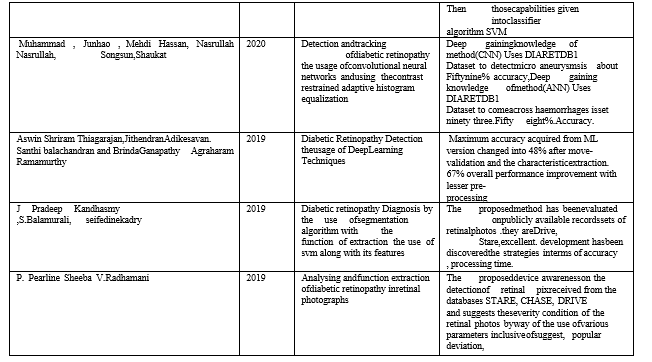
- In this paper studied the specific approximately the identification of DR inside the moderate nearly one hundred fifty studies articles, summarized with the gathering of retinal datasets, adoption of different forms of methodologies to find out the diabetic retinopathy and pick out the overall performance evaluation metrices for the instance in their consequences. Deep getting to know technique (CNN) Uses DIARETDB1 Dataset to come across the micro aneurysms is set ninety eight Fifty nine% accuracy, Deep gaining knowledge of approach (ANN) Uses DIARETDB1 Dataset to hit upon Exudates is set ninety eight% accuracy.
- In this paper sort of learning models were tried and the corresponding effects had been plotted. Random Forest models capture the underlying distribution of the snap shots in the information set fashions. The most accuracy obtained from ML model become forty-eight% after move-validation and the feature extraction. Sixty-seven% performance improvement with lesser pre-processing efforts, leaving the deep studying model a clear preference over the opposite traditional algorithm for automatic DR detection.
- In these studies, makes use of the furnished method allows to decreasing the manner area for segmentation techniques vital to increasing the general overall performance and reducing the amount of the desired computational price for each retinal fundus photograph. The proposed technique makes use of the available statistics gadgets of retinal photographs which is probably DIARETDB1, HRF, DRIVE, and a tremendous improvement decided in phrases of accuracy and processing time.
- In this paper uses the pre-processing and characteristic extraction of the diabetic retinal fundus picture for the diabetic retinopathy detection. Mean, trendy deviation, variance, imply squared mistakes and height sign to noise ratio gives specific price for category. The proposed machine collects the statistics for detection of retinal photos received from the STARE, CHASE, DRIVE
- In this paper makes use of diverse methods used to detect DR. Divide the technique into pre-processing, feature of DR detection, blood vessels segmentation, optic disc detection, EXs detection, MAs and HMs detection, and category the usage of SVM.Dataset collects the statistics from DRIVE, DIARETB0, DIARETB1, Mesidor, e-ophta-MA, e-ophta-EX.
- In this paper photo pre-processing is composed the Illumination Equalization, Denoising, Adaptive evaluation Equalization, and Colour Normalization. A new method used for pink lesion detection is proposed. The results of this approach give robust overall performance in detecting both microaneurysms and haemorrhages. The pre-processing steps which make lesions certainly seen.
- In this paper collects the input photograph statistics from DRIVE or STARE database. Uses Image pre-processing- photograph cropping, inexperienced channel extraction, assessment enhancement the usage of CLAHE, function extraction, neural network training, the skilled Neural network is examined for pics from the checking out database. The performance parameter given by way of sensitivity, specificity and accuracy and ROC suggests the higher normal performance values of the parameters.
- In this paper represents the 5-elegance problem for screening of diabetic retinopathy can be approached using a CNN method. CNN which makes a short analysis and instant response to a patient possible. Gives the accuracy quantity of patients with a correct type. Sooner or later trained community is completed about, 95% specificity, seventy-five% accuracy and 30% sensitivity.
- This paper offers the intelligence method for detecting initial lesions that seem inside the retina due to diabetic retinopathy. Colour fundus snap shots are considered as non- uniform illumination. Feature extraction and class gives the higher quit result for detection of lesion the usage of the digital fundus photographs. SVM is used for classifying and it provides better category accuracy even as compared to previous techniques.
- In this paper examine the pre-processing steps for retinal picture evaluation. Addressed the two issues of pre- processing of retinal images as noise removal and background extraction. Operations like Gaussian filtering, and morphological operations(average) and Top-Hat transformation were used for imperative mild reflex elimination, heritage extraction, blood vessels enhancement on retinal snap shots of publicly available DRIVE database.
III. COMPARISION TABLE
|
AUTHOR |
YEAR |
APROACH |
DESCRIPTION |
|
akshmi Narayanan, Khalafallah, Sarkar , Balaji |
2021 |
Analysis and detection of diabetic retinopathy |
DR detection are specifically targeted on automatic methods called CAD Systems Classification of DR may be divided into ML primarily based and DL primarily based. The evaluated DR proved that the DCNN architecture. |
|
Priyank Gandhi, Akshay, Govardhan, Shirish |
2021 |
Identify the Diabetic Retinopathy, the usage of Convolutional Neural Network |
Neural community used for image pre- processing , classifies the DR. Using the Confusion matrix to display the distribution of the photographs of fundus. |
|
M.Abirami, Vignesh, Vikram Sriram, E.Shivanithyesh. |
2021 |
Automatic Detection of diabetic retinopathy the usage of deep getting to know techniques |
CNN used for the automatic DR detection . Classify. The model for training the CNN reduces the complexity of the neural community. |
|
Abdelouahab Attia, Zahid , Samir, Sofiane maza. |
2020 |
Detection of diabetic retinopathy the use of system deep learning to know strategies |
Uses the retinal picture databases. Eight databases are to be had. DRIVE, STARE and Messidor dataset makes use of Exudates, haemorrhages, microaneurysms and extraordinary blood vessels detection. |
|
Azra Moment Pour, Hadi Seyedarabi, Seyed Hassan Abbasi Jahromi, AND Alizera Javadzadesh |
2020 |
CNN and evaluation and adaptive histogram equalization used to locate computerized detection of diabetic retinopathy |
Clache is used for the amplification of the blood vessels in retinal fundus pix because the image- processing step. Important features used to hit upon DR ranges. |
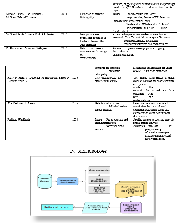
The Dataset is downloaded from the Kaggle website is used for pre-processing in order to standardize these image images ,reduce redundant information and environment artificats, several pre-processing methods such as filtering, and padding is applied.
- Convolutional Neural Network: Is also known CONVNet. The networks are multi-layer perceptron’s. The input fed to the model flows right through them so, these are called “feed-forward”. The model consists of five hidden layers and each layer performs three operations: Con-volution, Activation, and Pooling.
- Output: The prediction is expressed in the five forms are name No DR, Mild DR, Moderate DR, Severe DR and Proliferative.
Conclusion
Recognise the Diabetic Retinopathy the usage of retinal Fundus Images. Utilizing the deep learning of strategies within the diagnosis of a ailment based on retinal fundus pics, photo pre-processing, function extraction and classification used to identify the affected and non- affected a part of the retinal photograph. Deep Convolutional Neural Network is used to assemble our neural community fashions to classify retinal fundus snap shots amassed from the Kaggle internet site. Output the prediction expressed in five bureaucracy they\'re specifically Normal, Mild, Moderate, Severe and proliferative Diabetic Retinopathy.
References
[1] Lakshmi Narayanan, kheradfallah,H, Sarkar, Balaji.”Automatic detection and analysis of diabeticetinopathy”.2021 [2] Priyank, Akshay, Govardhan, Swapnil Jagtap.“Automatic Detection of Diabetic Retinopathy”,2021. [3] M.Abirami, M.Vignesh, okay.Vikramsriram, E.Shivanithyesh. “Diabetic Retinopathy Detection the usage of DeepLearning Techniques” ,2021. [4] Abdelouahab attia, Zahid , Samir, Sofiane maza. “A deep learning for detection of diabetic retinopathy”.2020. [5] Asra Moment Pour, Hadi Seyedarabi, Seyed Hassan Abbasi Jahromi, AND Alizera Javadzadesh . “Automatic detection and monitoring of diabetic retinopathy using convolutional neural networks and using the evaluation limited adaptive histogram equalization” 2020. [6] Muhammad , Junhao , Mehdi Hassan, Nasrullah Nasrullah, Song sun,Shaukat. “Detection and tracking of diabetic retinopathy the usage of convolutional neural networks and using the contrast restrained adaptive histogram equalization” 2020. [7] Aswin Shriram Thiagarajan,jithendran Adikesavan. Santhi Balachandran and Brinda Ganapathy Agraharam Ramamurthy. “Diabetic Retinopathy Detection the usage of Deep Learning Techniques”. Journal of computer technology 2019. [8] J Pradeep Kandhasmy ,S.Balamurali, seifedine kadry. “Diabetic retinopathy Diagnosis by the use of segmentation algorithm with the function of extraction the use of svm along with its features”. 2019. [9] P. Pearline Sheeba V. Radhamani.” Analyzing and characteristic extraction of diabetic retinopathy in retinal pix”. 2019. [10] Nisha., Panchal, Dr Darshak , Thakore, Dr.Tanmay ”Diabetic Retinopathy Detection”2018. [11] Ms.SheetalMarutiChougule, Prof. Renke”New picture Pre-processing approach in Di abetic Retinopathy ”.2017. [12] Dr. Kulwinder S Mann and Sukhpreet. “Retinal blood vessels segmentation the usage of synthetic neural networks for detection of diabetic retinopathy”.2017. [13] Harry Pratt, Frans , Deborach Broadbenf, Simon P Harding, Yalin.“CNN for Diabetic Retinopathy”.2016. [14] C.P.Reshma Chand, Dheeba. “Detection of Exudates in Retinal Colour Fundus Images”. 2015 [15] V.P. Patil, Wankhede.” Image Pre-Processing and segmentation steps for retinal blood vessels”.2014.
Copyright
Copyright © 2022 Mr. Anand M, Sanjana B Raj, Chinmayi K S, Harshitha S. This is an open access article distributed under the Creative Commons Attribution License, which permits unrestricted use, distribution, and reproduction in any medium, provided the original work is properly cited.

Download Paper
Paper Id : IJRASET44594
Publish Date : 2022-06-20
ISSN : 2321-9653
Publisher Name : IJRASET
DOI Link : Click Here
 Submit Paper Online