

Ijraset Journal For Research in Applied Science and Engineering Technology
A Survey Paper on Pneumonia Detection in Chest X-Ray Images Using an Ensemble of Deep Learning
Authors: Dr. Manishankar S, Gowravi Sumana M, Khyathi Jain, Sneha C
DOI Link: https://doi.org/10.22214/ijraset.2022.44569
Certificate: View Certificate
Abstract
Pneumonia is a transferable infection influencing one or the two lungs in people ordinarily brought about by microorganisms called Streptococcus pneumonia. Chest X-Rays that are wont to analyze pneumonia need master radiotherapists for assessment. In this way, fostering a programmed framework for identifying pneumonia would be valuable. Convolutional Neural Networks (CNNs) certainly stand out enough to be noticed for infection arrangement involving profound learning calculations in investigating clinical pictures. Also, highlights advanced by pre- prepared CNN models for huge scope datasets from Kaggle are a lot helpful in picture order errands. during this work, we evaluate the usefulness of pre-prepared CNN models used as component extractors followed by various classifiers for the arrangement of strange and ordinary chest X-Rays. We scientifically decide the best possible Convolution Neural Network model for the point. Measurable outcomes acquired show that pre-prepared CNN models utilized along with directed classifier calculations are frequently exceptionally helpful in breaking down chest X-ray pictures, explicitly to distinguish Pneumonia.
Introduction
I. INTRODUCTION
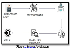
Compelling and precise medical care has forever been the need of great importance. Early recognition of changed infections like pneumonia, growth, and disease is altogether fundamental. Pneumonia, an intense respiratory lot disease positioned eighth on the rundown of the greatest 10 reasons for death in us. per WHO, it represents around 1.6 million passings every year during this accomplice - 18% of all passings among kids under five. The sickness habitually goes disregarded and untreated until it's arrived at a lethal point, particularly on account of old patients. A Series of tests like a Pleural liquid test, Sputum test, blood test, CT check, Pulse oximetry, and Chest X-ray are led for the determination which moves slowly. Chest X-rays are principally utilized for the conclusion of this sickness. Be that as it may, in any event, for a prepared radiologist, it's a provoking undertaking to check out at chest X-rays. In this venture, we construct an AI model, which takes X-ray pictures as information then continues to perform different picture handling tasks to recognize the locale of disease and train the convolution brain organization (CNN) model to group and distinguish the presence of pneumonia from an assemble of chest X-ray picture tests. This model could assist with relieving the dependability and interpretability challenges frequently confronted while overseeing clinical symbolism.
II. RELATED WORKS
- In the essential paper, we must be constrained to comprehend that there's a straightforward relationship between the finding of pneumonia and irregularities on CXR. Hence, applying profound figuring out how to discover pneumonia is a powerful symptomatic arrangement. Besides, this paper applied an outfit model utilizing Mask R-CNN and RetinaNet to RSNA Pneumonia Detection Challenge on Kaggle and shows the way that this technique can accomplish high precision of expectation. Since there's a significant development in picture handling regions like grouping and article identification, Convolutional Neural Network is typically utilized in programmed analysis with clinical pictures. the preeminent compelling much obliged for diagnosing pneumonia are irregularity discovery in Chest X-rays pictures. Pneumonia patient's CXR.
- The improved Mask RCNN model is achieved by building pneumonia recognition models under varied spine organizations in this future research. Models for detecting pneumonia are RetinaNet and Mask R-CNN. This research evaluated a technique on a collection of 26,684 radiographs from Kaggle, attaining a par of 0.813 and an mAP of 0.2283. Pneumonia is a disease brought about by infections, microscopic organisms, or different microorganisms. With the improvement of man-made brainpower toward PC vision, involving existing specialized means and programming for analysis is extremely dynamic and the objective discovery in clinical imaging is principally to tackle the issue of target positionings, like obsessive situating, atomic location, and strange point recognition. This piece will address the topic of pneumonia in chest X-rays. It essentially presents an approach to determining pneumonia using a set of diverse systems RetinaNet and Mask R-CNN.
- Through this paper, we advance completely about how pneumonia causes extreme impacts on people. Pneumonia affects around 7% of the whole population, with 4 million individuals in danger of mortality. Chest X-rays used to diagnose pneumonia require the expertise of professional radiotherapists. In this way, cultivating a pre-programmed architecture for recognizing pneumonia would indeed be beneficial in curing the infection with no postponement, especially in far-off regions.. Due to the progress of profound learning calculations in dissecting clinical pictures, Convolutional Neural Networks stand out for illness order. Measurable outcomes got to show that pre-prepared CNN models utilized along with directed classifier calculations might be exceptionally valuable in breaking down chest X-ray pictures, explicitly to distinguish Pneumonia. The grouping including high-rich deleted highlights demonstrates improved execution in arranging images. X-rays are regarded as the first successful method of determining the extent and condition of the infectious district inside the respiratory system in the diagnosis of pneumonia. In the X-ray pictures, the vibes of pneumonia are frequently cloudy and may be confused with different analyses. The assessment of chest X-Ray explicitly inside the instance of Pneumonia might be deceiving in light of the fact that numerous different issues like a congestive coronary disappointment, lung scarring, and so forth can mirror Pneumonia. The display of several permutations of convenient CNN models describes a description of the various classes for distinguishing between extraordinary and conventional chest X-Rays.
- This research depicts the utilization of AI calculations to handle chest X-ray pictures to help the dynamic cycle in deciding the right finding. In particular, the exploration is centered around the utilization of profound learning calculations in view of a convolutional brain organization to fabricate a handling design. This model is tasked with assisting with such a characterization challenge, which is determining if a chest X-ray exhibits change consistent with pneumonia and classifying the pictures into two gatherings relying upon the identification findings.
- In this paper, five distinct profound learning models,i.e, ResNet18, ResNet34, InceptionV3, InceptionResNetV2, and DenseNet161 and their Ensemble are utilized to arrange COVID-19, pneumonia, and solid prime diseases utilizing Chest X-Ray pictures. Multi-name characterization was conducted to foresee numerous pathologies for each understanding if present here. Premier, the in tractability of all of the organizations was totally concentrated on utilizing methods like impediment, saliency, input X angle, directed backpropagation, coordinated inclinations, and DeepLIFT. The mean Micro-F1 score of the models for COVID-19 orders goes from 0.66 to 0.875 and is 0.89 for the totality of the organization models. The subjective outcomes portrayed the ResNets to be the principal explicable models.
- This paper presents a model upheld DeepConv-DilatedNet for recognizing and limiting pneumonia in CXR pictures. Two-stage identifier Faster R-CNN is embraced due to the construction of an organization. Include Pyramid Network (FPN) is incorporated to the remaining brain organization of an enlarged bottleneck so the profound elements are extended to safeguard the profound component and position data of the article. Inside the instance of DeepConv-DilatedNet, the network is utilized to resuscitate undeniable level component guides to their unique size, and accordingly the objective data is additionally held. On the contrary hand, DeepConv- DilatedNet utilizes a well known completely convolution design with calculation shared in general picture. Then, at that point, Soft-NMS is utilized and guarantee test quality. Additionally, K-Means++ is utilized to concoct anchor boxes to improve restriction precision. The calculation acquired 39.23% Mean Average Precision on the X-ray picture dataset from the Radiological Society of North America and got 38.02% Mean Average Precision on the ChestX-ray14 dataset, outperforming other discovery calculations.
- This research centers around studying and contrasting the location of lung infection utilizing different PC-supported methods and proposes a reexamined model for identifying pneumonia. Here they likewise attempted to get to know the different picture pre-handling procedures wont to change over crude CXR pictures into standard configurations for examination and recognition, AI strategies likeKNN, RESNET, Chenet, CNN, ANN, and DENSENET, which is an indispensable stage in exact infection location. Pre-handling strategies for the picture are utilized here to erase unimportant information. Leveling of the histogram works on the picture and sifting of the picture lessens commotion and hones the picture in a high pass channel. Area of interest is utilized for the production of lung division. Demonstrative elements like edge, region, anomaly file, and equivalent measurement and inconsistency record are removed on well as factual highlights like change, mean, and entropy. Feed-forward and back-spread brain networks are utilized for picture characterization to identify lung illnesses.
- This exploration portrays the work of AI calculations to handle chest X-ray pictures to help the dynamic interaction in deciding the right analysis. In particular, the exploration is designated at the usage of profound learning calculations upheld by a convolutional brain organization to construct a handling model. This model's task is to assist with an arrangement issue that is determining if a chest X-ray reveals change in regards to pneumonia or not and classifying the X- ray photos into two groups based on the discovery outcomes
- In this paper, we present a brand new chest X-ray data base, specifically "ChestX-ray8," which contains 108,948 front facing view X-ray pictures of 32,717 unique patients with message mined eight illness picture names (where each picture can have multiple marks), and extracted from related radiological reports utilising language handling. We show that these often occurring thoracic diseases may be detected and, unexpectedly, geographically discovered using a bound together feebly administered multi-mark image characterisation and infection confinement structure, which is validated using our suggested dataset. ] Despite the fact that the underlying quantitative outcomes are promising, deep convolutional brain network-based "reading chest X-rays" (i.e., perceiving and finding the typical infection designs prepared with just picture level names) remains a difficult task for completely computerised high accuracy CAD frameworks.
- The job of numerous tiered convolutional network (HCN) engineering is proposed in this study to usually enlarge the data together with distinct highlights. To extract the highlights, the HCN employs the fundamental convolution layer from COVIDNet, followed by convolutional layers from well-known pre-prepared organisations. The use of the COVIDNet convolution layer ensures the extraction of depictions suitable for the CXR approach. We also recommend using ECOC for encoding multiclass concerns to twofold grouping in order to improve acknowledgment execution. Exploratory outcomes show that HCN design is equipped for accomplishing improved outcomes in examination with the current investigations. The proposed technique can precisely emergency potential patients through CXR for sharing the testing load and expanding the limit.
- This paper proposes a mass identification model upheld RetinaNet. To approve its presentation in assorted use cases, we develop a few trial arrangements utilizing the public dataset INbreast and furthermore the in-house dataset GURO. As well as preparing and testing on the indistinguishable dataset (i.e., preparing and testing on INbreast), we assess our mass discovery model in arrangements utilizing extra preparation information (i.e., preparing on INbreast + GURO and testing on INbreast). We additionally assess our model in arrangements utilizing pre- prepared loads (i.e., utilizing loads pre-prepared on GURO, preparing, and testing on INbreast). by and large the analyses, our mass location model accomplishes practically identical or preferable execution over more complicated cutting edge models including the two-stage object finder. Likewise, the outcomes show that utilizing the loads pre-prepared on datasets accomplishes comparable execution as straightforwardly utilizing datasets inside the preparation stage. In this way, we make our mass recognition model's loads pre-prepared on both GURO and INbreast openly accessible. We expect that analysts who train RetinaNet on their in-house dataset for the mass location assignment can utilize our pre-prepared loads to use the elements extricated from the datasets.
- In this paper, the primary spotlight is on summing up the best in class works related with profound applications for COVID-19 clinical picture handling. With the steady movement after some time, COVID-19 was pronounced by the planet wellbeing association (WHO) as an endemic, which has forced a huge weight on most nations, particularly ones with more vulnerable wellbeing frameworks and ones with slow reactions. inside the field of medical care, profound learning been has carried out in numerous application.Various wellsprings of clinical pictures make profound learning an extraordinary method to battle the COVID-19 episode Then, it gives a diagram of profound learning and its applications to medical care found inside the last 10 years. Then, three use cases in China, Korea, and Canada additionally are introduced to demonstrate applications for COVID-19 clinical picture handling. At last, it talks about a few difficulties and problems related with profound learning executions clinical picture handling, which are supposed to guide further examinations in controlling the episode and the emergency, which closes in savvy solid urban areas.
- In this article, pre-prepared Convolutional Neural Networks (CNN) on chest x-ray images are used as component extractors, which are subsequently used to describe the images to predict whether someone has pneumonia. In terms of forecasting on images, the various pre-prepared Convolutional Neural Networks used are tested with varied bounds. The impacts of pre-prepared brain networks were investigated, and a troupe model was proposed that combines the forecasts of the least challenging pre-prepared models to provide better results than individual models.
- In this paper, we concentrate on certain approaches to melding ConvNet towers spatially and transiently to the best benefit from this Spatio-fleeting data. We make the resulting discoveries: (I) that as opposed to combining at the softmax layer is a spatial and worldly organization that will be intertwined at a convolution layer, yet with an impressive saving in perimeter; (ii) that it's smarter to circuit such organizations spatially at the layer than prior, which also melding at the classification forecast layer can support precision; at last (iii) pooling of dynamic convolutional highlights over Spatio-transient areas again lifts execution. upheld these examinations we propose another ConvNet engineering for the Spatio-worldly combination of video scraps, and assess its exhibition on standard benchmarks where this design accomplishes cutting edge outcomes.
15. In this paper,a profound learning structure is recommended that coordinates a convolutional brain organization and a case organization. DenseCapsNet, a pristine profound learning system, is made by the combination of a thick convolutional network (DenseNet) and in this manner the container brain organization (CapsNet), utilizing their separate benefits and lessening the reliance of convolutional brain networks on an outsized measure of dataset. Utilizing 750 CXR pictures of lungs of sound patients additionally as those of patients with other pneumonia and novel Covid pneumonia, the system can get an exactness of 90.7% and a F1 score of 90.9%, and in this way the awareness for recognizing COVID-19 can reach 96%. These outcomes show that the profound combination brain network DenseCapsNet has great execution in novel Covid pneumonia CXR radiography location
III. COMPARISION TABLE
|
AUTHOR |
YEAR |
APPROACH |
DESCRIPTION |
|
Shangjie Yao ,Yaowu Chen ,Xiang Tian and RongxinJiang3 |
2018 |
DeepConvDilated Net K-Means++ CLAHE Algorithm Soft- NMSalgorithm |
Consolidating the contrasting arrangements of work depleted in each and every organization, The effort of the calculation to identify pneumonia precisely inside the RSNA is improved. |
|
Xianghong Gu, Liyan Pan, Huiying Liang,Ran Yang |
2018 |
A deep convolutional neural network (DCN) is a type of neural network that In chest radiography, a CAD system to detect bacterial and viral pneumonia. |
The technique comprises of two sections, lung districts recognizable proof,and pneumonia classification arrangement |
|
Benjamin Antin, JoshuaKravitz, and Emil Martayan |
2019 |
CheXNet 121- layerdense Convolutional Neural Network |
The technique comprises of two sections,lung districts recognizable proof,and pneumonia classification arrangement |
|
Xiaowei,XuXian gao,JiangChunli a n MaPeng ,DuXukun |
2020 |
Noisy-OR Bayesian function a three- dimensional CNN segmentation model |
Two arrangement models are utilized; one is a somewhat conventional leftover organization and other is planned in light of the intial network structure by joining the location consideration mechanism to work on the by and large precision rate. |
|
Soumick Chatterjee,Fatima Saad,Chompu nuch Sarasaen, Suhita Ghosh |
2020 |
ResNet18, ResNet34, InceptionV3, InceptionResNet V2, and DenseNet16 |
Prior non-picture information can additionally be attempted to be integrated into the organization models |
|
Saurabh Vernekar Dhanashri Shrameet Nayak Turi Pratiksha R. Shetgaonkar Ashitosh Tilve |
2020 |
CNN, Residual neural network, CheXNet, DenseNET |
It can be shown thatVGG16 accomplishes the most elevated exactness, implying that different image preprocessing approaches can increase the organization's speed and precision. |
|
Sweta Bhattachara , Praveen Kumar Reddy Maddikunta,Thi ppaReddy Gadekallu , Siva Rama Krishnan S , Chiranji Lal Chowdhary , Mamoun Alazab , Md. Jalil Piran |
2020 |
ML models, CNN using triple cross- validation |
DL has long been regarded as a valuable tool for developing intelligent solutions. We have detailed current work regarding the COVID19 epidemic for smart, healthy cities, motivated by the preceding decade, there were various applications of DL for computer-aided diagnosis. |
|
Luka Racic, Tomo Popovic, Stevan Cakic, Stevan Sandi |
2020 |
Machine learning algorithm of CNN |
Dropout is a mechanism in which certain neurons are turned off at random and are not used in the next iteration. The network can improve its accuracy by 1-2 percent just by adding a dropout. |
|
Yuechun Shen1,7, YuelinChen1,2, 7, Zheng Huang1, Junyao Huang3, Xinchun Li 4, ZuojunTian5 & JunLi 6 |
2020 |
Excel sofware. SPSS sofware (Version 17; SPSS, Inc) Mean±stand ard deviation |
Excel software was wont to manage the info. SPSS software (Version 17; SPSS, Inc) was wont to perform statistical analyses. Mean±standard deviation was accustomed express continuous variables. Percentage or number was wont toexpress categorical variables. |
|
V S Suryaa, Arockia Xavier Annie R, Aiswarya M S |
2021 |
DenseNet Architecture, VGG Architecture, MobileNetV2 Architecture |
Pneumonia identification using chest X-rays is automated using an ensemble model. To propose an ensemble mode, various CNN structures were fine-tuned, trained, and the obtained results were examined. |
IV. METHODOLOGY
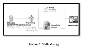
- Dataset: The dataset used is Chest X-ray which is publicly to be had on the Kaggle33platform which consists of a complete of 5836 pics segmented into two main parts, a schooling set and a check set. Both bacterial and viral pneumonia are considered as a unmarried category, pneumonia infected. The entire chest X-ray imaging turned into performed as a part of patients’ recurring medical care. Each image needs to be preprocessed in line with the deep neural network used.
- Preprocessing: Each image had to be preprocessed in step with the deep neural community used. The critical steps involved are: resizing and normalization. Different neural networks require photos of various sizes in keeping with their described structure. All the images are to be normalized according to the respective architectures.
- Data Augmentation: The facts is imbalanced i.e., the X-ray images of the non-pneumonia elegance are more in comparison to the pneumonia X-ray photographs. Data augmentation would be carried out on our X-ray photograph i.e., artificially make bigger the scale of our high-quality magnificence (X-ray images with pneumonia). Many information augmentation techniques consist of inclusive of Horizontal and Vertical Shift Augmentation, Horizontal and Vertical Flip Augmentation, Random Rotation Augmentation, Random Brightness Augmentation and plenty of greater. Model Convolution neural networks (CNNs) belong to a category of Deep Learning models that are especially used within the discipline of computer vision. These models have multiple processing layers to learn hierarchical function representations from the input pixel facts. It allows in learning the model for extracting the features after which constructs the item for classifying the venture. CNN models are feed-forward networks with convolution layers, pooling layers, knocking down layers and completely connected layers using suitable activation features. The principal benefit of CNN is that it's miles able to detecting the applicable functions without any human supervision.
- User side: GUI User side offline model interface would be built, standard GUI library for Python. The users have to upload the X-ray.
Conclusion
The review describes an exchange knowledge group model for automating Pneumonia recognition utilizing Chest X-rays. Various CNN structures were tweaked and created, and the results were dissected to eventually suggest a group model. The second fundamental idea focused on the production is to reduce the purpose and size of the images used while compensating the compromise with the model\'s exposition. In reality, such models may be given to reduce doctors\' responsibilities and reduce human error levels. With a smaller capacity restriction and processing talented specialists and radiologists, unfortunate network, and absence of foundation. Even though it can\'t supplant a doctor, it can help the finding system and lessen the essential time taken.
References
[1] Pneumonia Detection Using an Improved Algorithm Based on Faster R-CNN by Shangjie Yao, Yaowu Chen, Xiang Tian, and Rongxin Jiang,Japan,2018 [2] Classification of Bacterial and Viral Childhood Pneumonia by Using Deep Learning in Chest Radiography byXianghong Gu, Liyan Pan, [3] Huiying Liang, Ran Yang,, Springer, 2018 [4] Detecting Pneumonia in Chest X-Rays with Supervised Learning by Benjamin Antin, Joshua Kravitz, and Emil Martayan, Barcelona, [5] Spain,2019 [6] A Deep Learning System to Screen Novel Coronavirus Disease2019PneumoniabyXiaowei,XuXiangao,JiangChunlian MaPeng [7] DuXukun ,Japan,2020. [8] Exploration of interpretability techniques for deep covid-19 classification using chest x-ray images by Soumick Chatterjee, Fatima Saad, Chompunuch Sarasaen, Suhita Ghosh, Germany,2020 [9] A. Mangal, S. Kalia, H. Rajgopal et al., “CovidAID: COVID-19 detection using chest X-ray,” 2020 [10] T. Ozturk, M. Talo, E. A. Yildirim, U. B. Baloglu, O. Yildirim, and [11] U. R. Acharya, “Automated detection of COVID-19 cases using deep neural networks with X-ray images,” Computers in Biology and Medicine, 2020. [12] [8]C. F. Kuo and H. C. Wu, “Gaussian probability bi-histogram equalization for enhancement of the pathological features in medical images,” International Journal of Imaging Systems and Technology 2020. [13] L. Wang and A. Wong, “COVID-Net: a tailored deep convolutional neural network design for detection of COVID-19 cases from chest X- ray images,” 2020. [14] D. L. Donoho, “Compressed sensing,” IEEE Transactions on Information Theory. [15] R. G. Baraniuk, “Compressive sensing,” IEEE Signal Processing Magazine, vol. 24, no. 4, pp. 118–120, 2017 [16] M. Fiszman, W. W. Chapman, S. R. Evans, and P. J. Haug, “Automatic identification of pneumonia-related concepts on chest x-ray reports.,” in Proc. of the AMIA Symposium, p. 67, American Medical Informatics Association, 2019. [17] W. W. Chapman, M. Fizman, B. E. Chapman, and P. J. Haug, “A comparison of classification algorithms to automatically identify chest x- ray reports that support pneumonia,” Journal of Biomedical Informatics, vol. 34, no. 1, pp. 4–14 [18] P. Rajpurkar, J. Irvin, K. Zhu, B. Yang, H. Mehta, T. Duan, D. Ding, A. Bagul, C. Langlotz, K. Shpanskaya, et al., “Chexnet: Radiologistlevel pneumonia detection on chest x-rays with deep learning,”. [19] E. A. Mendonca, J. Haas, L. Shagina, E. Larson, and C. Friedman, “Extracting information on pneumonia in infants using natural language processing of radiology reports,” Journal of Biomedical Informatics. [20] gadget requirements, the execution might be brought to far provincial regions internationally that need legitimate findings and treatment for such sicknesses because of the absence of
Copyright
Copyright © 2022 Dr. Manishankar S, Gowravi Sumana M, Khyathi Jain, Sneha C. This is an open access article distributed under the Creative Commons Attribution License, which permits unrestricted use, distribution, and reproduction in any medium, provided the original work is properly cited.

Download Paper
Paper Id : IJRASET44569
Publish Date : 2022-06-19
ISSN : 2321-9653
Publisher Name : IJRASET
DOI Link : Click Here
 Submit Paper Online