

Ijraset Journal For Research in Applied Science and Engineering Technology
Diagnosis of Diabetic Retinopathy
Authors: Pawan Sharnagate, Pooja Nimje, Mukul Bisen , Kankeshwari Baywar, Ashish Raj, Prof. Ms. Minakshee Chandankhede
DOI Link: https://doi.org/10.22214/ijraset.2022.48121
Certificate: View Certificate
Abstract
A diabetes-related eye condition called diabetic retinopathy (DR) can cause blindness or blurred vision. The risk of vision loss can be significantly reduced with early DR diagnosis and treatment. Due to the heterogeneity of retinal alterations between disease stages and the significant proportion of misdiagnosed and untreated DR cases, such diagnosis is a time-consuming and challenging endeavour. A transfer learning method is used to speed up the process while several preprocessing algorithms are used to increase accuracy. The fundus image collection from the online Kaggle datasets was used in our experiment.
Introduction
I. INTRODUCTION
A. Overview
|
|
Diabetic retinopathy (DR) is recognized as one of the world's leading blinding diseases, especially in the elderly.
In this work, we present an intelligent system for DR classification based on deep learning (DL) tools, specifically convolutional neural networks (CNNs).
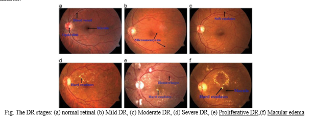
The proposed system helps ophthalmologists make preliminary decisions and enables DR classification considering normal eye, mild DR, moderate DR, severe DR and proliferative DR .
Images based on VGG-16 and ResNet50 models are 70% and 25%, respectively.
However, the diagnosis of DR is a tedious and complicated task due to the variability of retinal changes in disease stage and the large number of undiagnosed and untreated DR cases.
- Stage 1: Mild nonproliferative diabetic retinopathy (0,1)
This is the earliest stage of diabetic retinopathy, characterized by tiny areas of swelling in the blood vessels of the retina. These areas of swelling are known as micro aneurysms.
2. Stage 2: Moderate nonproliferative diabetic retinopathy (0,1,2)
Increased swelling of tiny blood vessels starts to interfere with blood flow to the retina,
3. Stage 3: Severe nonproliferative diabetic retinopathy (0,1,2,3)
A larger section of blood vessels in the retina become blocked, causing a significant decrease in blood flow to this area.
4. Stage 4: Proliferative diabetic retinopathy (0,1,2,3,4)
This is an advanced stage of the disease, in which new blood vessels form in the retina. Since these blood vessels are often fragile, there’s a higher risk of fluid leakage. This triggers different vision problems such as blurriness, reduced field of vision, and even blindness.
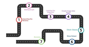
II. METHODOLOGY
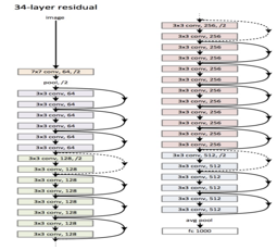
Based on our dataset, each Deep Learning, CNN (Convolutional Neural Network) and ResNet (ResNet-34) were trained and tested with many refinements for each model. Resnet34 is a 34-layer convolutional neural network that can be used as a state-of-the-art image classification model. This is a model pre-trained on the ImageNet dataset, a dataset containing over 100,000 images in 200 different classes. The traditional ResNet-34 model contains 63.5 million parameters, with Rectification Nonlinearity Activation (ReLU) and Batch Normalization (BN) applied behind every convolutional layer.
A. ResNet-34
Resnet34 is a 34 layer convolutional neural network that can be utilized as a state-of-the-art image classification model. However, it is different from traditional neural networks in the sense that it takes residuals from each layer and uses them in the subsequent connected layers . This is because when the network is too deep, the gradients from where the loss function is calculated easily shrink to zero after several applications of the chain rule. ResNet-34/DR is composed of five Convolutional groups in each group, where one or more Conv layer output passes through the BN layer and ReLU As a sequence (Conv→ BN→ ReLU) .This result on the weights never updating its values and therefore, no learning is being performed.
III. IMPLEMENTATION
A. Image Filtering
Images are created using different camera resolutions, different sizes, and a variety of effects. They are unprocessed data. To reduce noise and improve the accuracy of our model, these data were incorporated into the image processing process. Ben Graham's method served as a model for this treatment and was modified. Although it employs different kernels to produce Gaussian fragments, it is comparable to medianfiltering. To eliminate noise from such photos, this is done. The final stage involves cropping the image's outer 10% of its perimeter on both sides, which removes all of its important information..
B. Data Augmentation
More data during deep learning model training can result in more intelligent models. Data augmentation enhances the generalizability of the model and lessens the impacts of overfitting. The best data augmentation technique depends a lot on the nature of the issue, though. In this study, we present a data augmentation and image enhancement technique for the classification of pigmented macular anomalies in non-mydriatic fundus pictures. With the aid of several processing techniques or a combination of multiple different processing techniques, such as random rotation, resizing, mirroring, cropping, and flipping, a fake training image is created.
IV. TRAINING
The validation set examines the development in model performance over time and chooses the optimal parameter after splitting the dataset into training and validation sets in the training phase. If the training accuracy does not improve after ten consecutive cycles or the validation set performs worse, an early stopping method was utilised to end training. If training is terminated early and the validation error is not recovered, optimal weights are retained.
Conclusion
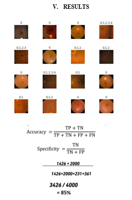
ResNet 34 Convolution Neural Networks has been shown that have high potential and accurate results to be training for detection of the features of DR in the APTOS images and to the automated retinal images analysis. This project successfully detects diabetics by using deep learning with 85% accuracy and save time and cost at a same time. Improvements in diabetes and diabetic retinopathy treatments have resulted from better understanding of pathophysiology and clinical trials that show the benefits of aggressive approaches .Improvements will build on these successes to further reduce the risk of vision loss and will lead to early diagnosis and less invasive treatments. When prediction of diseases is automated, time will be minimized so that people can take preventive measures beforehand. In the future the parameters of the algorithms can be finely tuned to get better results and the accuracy of the model can be improved using other efficient optimization techniques.The project can be enhanced with the user interface implementation for making it available in real time for the users. This would ease the burden of the doctors.
References
[1] Benbassat, J., Polak, B.C.. Reliability of screening methods for diabetic retinopathy. Diabetic Med 2009;26(8):783–790. [2] Grading diabetic retinopathy from stereoscopic color fundus photographsan extension of the modified airlie house classification: Etdrs report Number 10. Ophthalmology 1991;98(5):786–806. [3] Philip, S., Fleming, A.D., Goatman, K.A., Fonseca, S., Mcnamee, P., Scotland, G.S., et al. The efficacy of automated disease/no disease Grading for diabetic retinopathy in a systematic screening programme. Brit J Ophthalmol 2007;91(11):1512–1517. [4] Fleming, A.D., Philip, S., Goatman, K.A., Prescott, G.J., Sharp, P.F., Olson, J.A.. The evidence for automated grading in diabetic Retinopathy screening. Current Diabetes Reviews 2011;7:246 – 252. [5] Mookiah, M.R.K., Acharya, U.R., Chua, C.K., Lim, C.M., Ng, E., Laude, A.. Computer-aided diagnosis of diabetic retinopathy: A Review. Comput Biol Med 2013;43(12):2136–2155. [6] Fukushima, K.. Neocognitron: A self-organizing neural network model for a mechanism of pattern recognition unaffected by shift in Position. Biol Cybern 1980;36(4):193–202. [7] Srivastava, N.; Hinton, G.; Krizhevsky, A.; Sutskever, I.; Salakhutdinov, R. Dropout: A Simple Way to Prevent Neural Networks from Overfitting. J. Mach. Learn. Res. 2014, 15, 1929–1958. [8] Chollet, F. Deep Learning with Python, 1st ed.; Manning Publications Co.: Greenwich, CT, USA, 2017;ISBN 9781617294433.
Copyright
Copyright © 2022 Pawan Sharnagate, Pooja Nimje, Mukul Bisen , Kankeshwari Baywar, Ashish Raj, Prof. Ms. Minakshee Chandankhede . This is an open access article distributed under the Creative Commons Attribution License, which permits unrestricted use, distribution, and reproduction in any medium, provided the original work is properly cited.

Download Paper
Paper Id : IJRASET48121
Publish Date : 2022-12-13
ISSN : 2321-9653
Publisher Name : IJRASET
DOI Link : Click Here
 Submit Paper Online